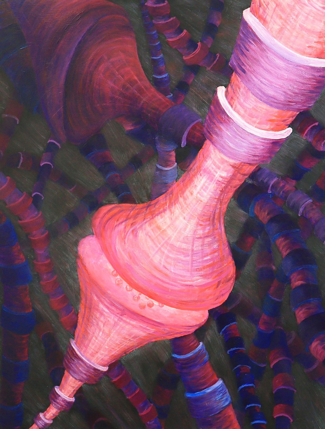
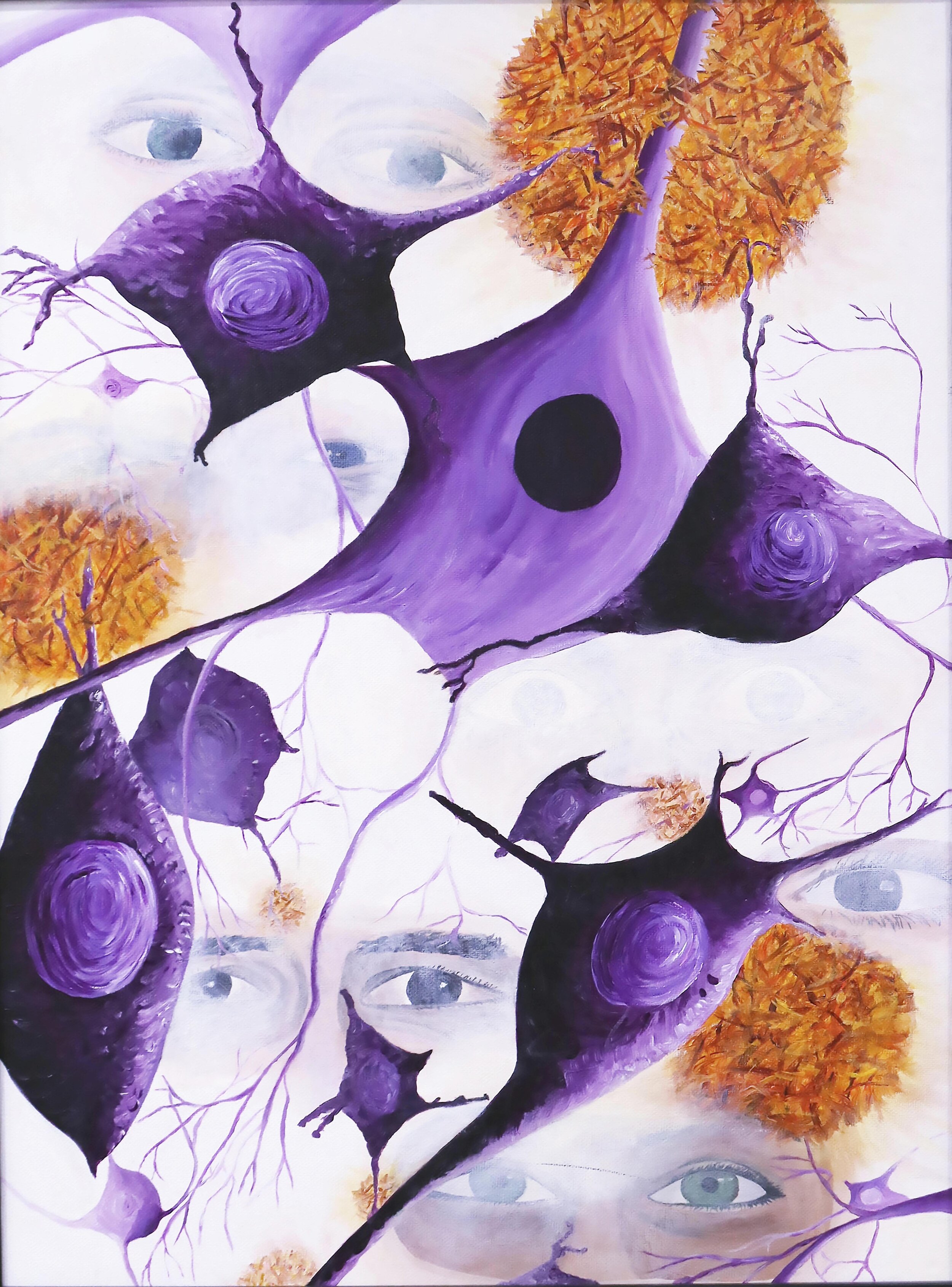
18×25” acrylic on wood
A synpase occurs between two neurons and allows brain cells to send messages to each other. The axon of one neuron sends an electrical current to the end where it triggers release of chemicals, called neurotransmitters, which diffiuse across a gap to the dendrite of another neuron. These chemical bind specific receptors and pass the message along converting it back to an electrical signal.
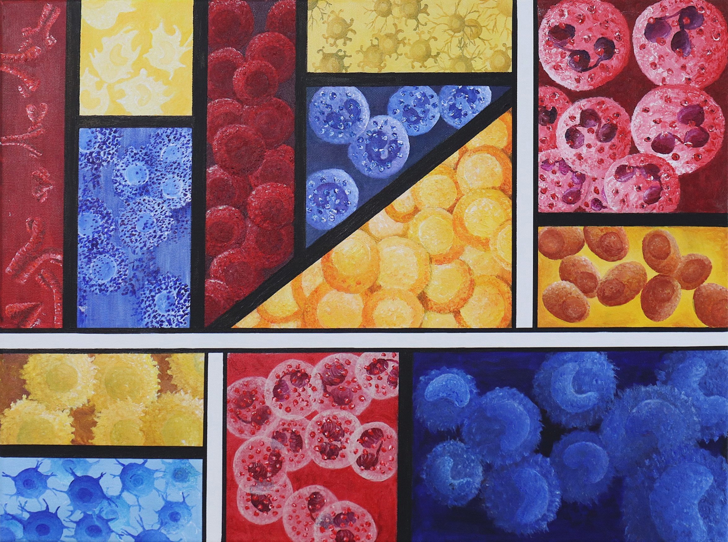
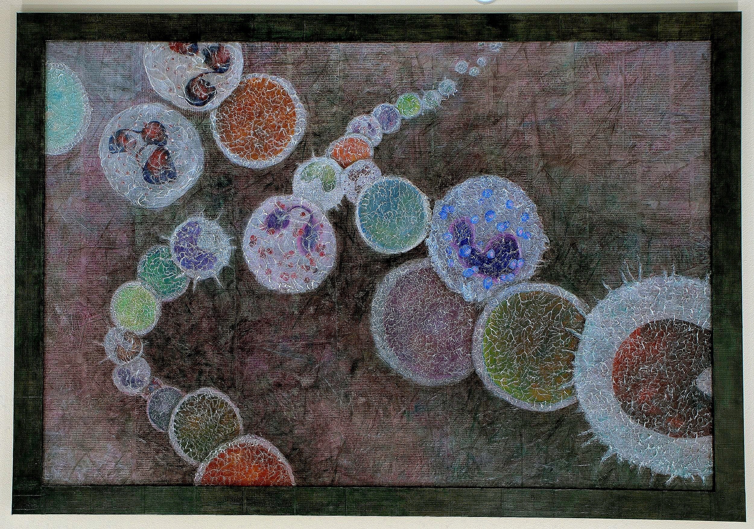
18 ‘×24” Acrylic on Canvas
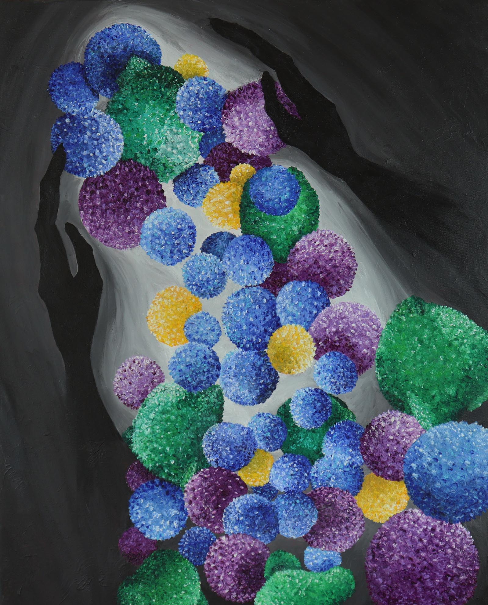
The immune system protects the body from infection, is important in the healing process, and plays a critical role in a variety of diseases. There are a variety of cells within the immune system that each play a unique role, and interact with each other to promote inflammation, healing and recovery. This painting, inspired by the work of Piet Mondrian, shows the diversity of cells found throughout the immune system. I used primary colors divided by white and black lines to create this abstract piece.

16×20” acrylic on canvas
Rheumatoid arthritis is an autoimmune disease that most commonly affects joints of the hands. Pain, swelling and stiffness are common, along with widespread fatigue. Current disease modifying drugs include antibody treatments aimed at modifying the immune system - these have transformed the course of the disease.

16×20” acrylic on canvas
Ion channels and receptors are found in the lipid membrane of cells including neurons. They are proteins that respond to stimuli by binding to their large external domain. Proteins are depicted though ribbon diagrams initially developed and hand drawn by Jane Richardson in 1980.
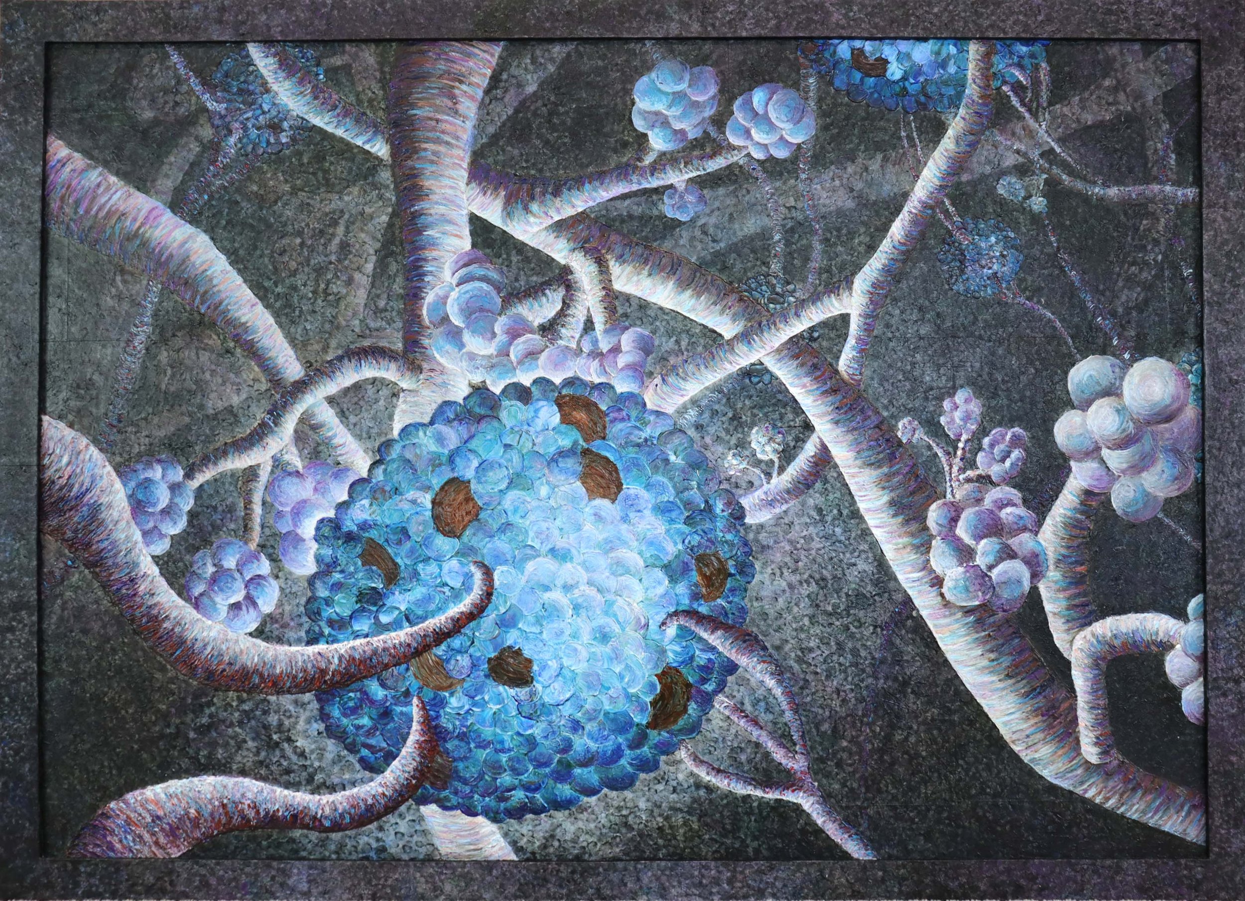
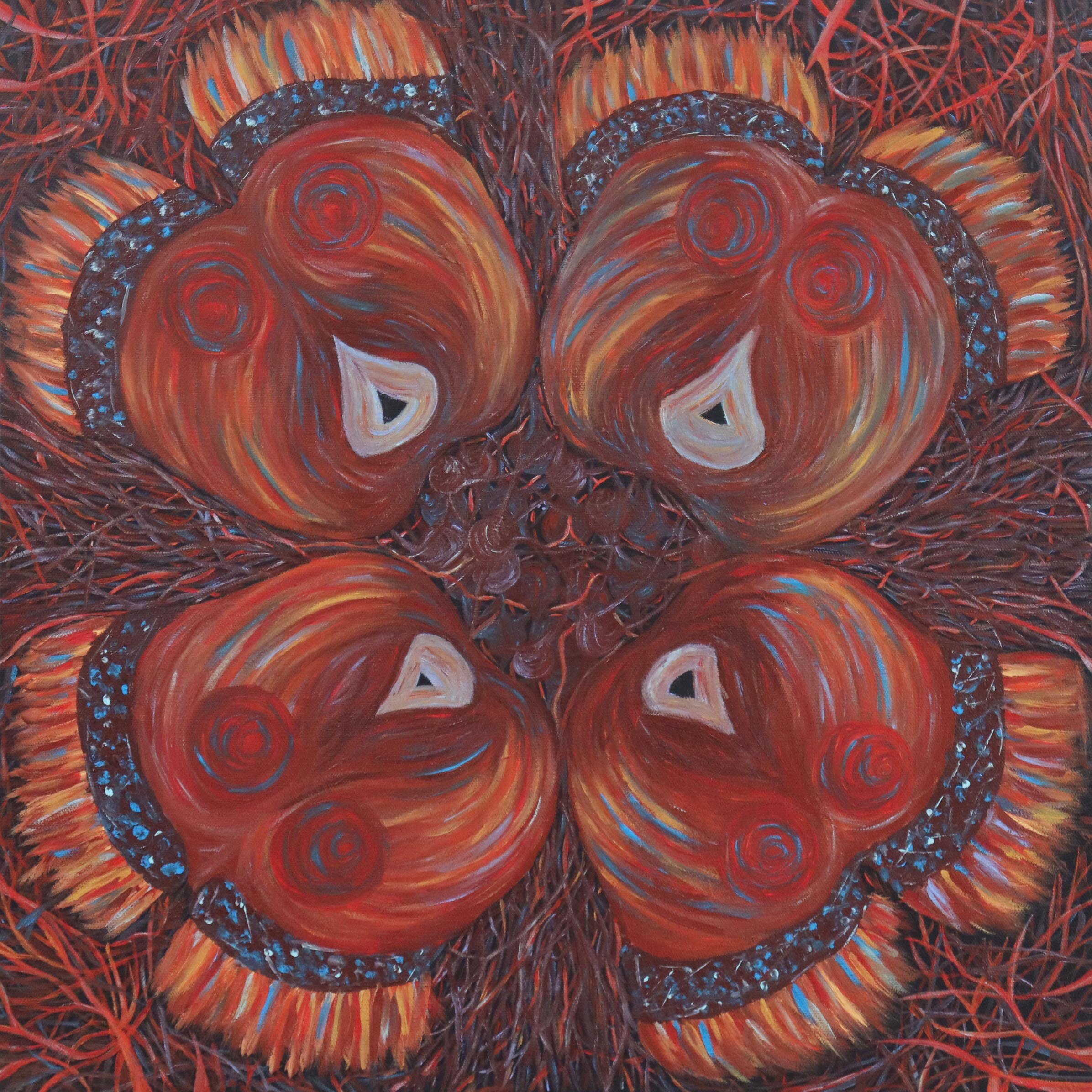
3 x 4 foot acrylic painting on wood
The pancreas is responsible for maintaining proper blood sugar levels. Islets of Langerhans contain two types of cells that produce insulin to decrease, and glucagon to decrease blood sugar levels. The pancreas also contains acinar (grape-like cells) that synthesizes and secretes digestive enzymes that are then released into the small intestine. Dysfunction in the islet cells can lead to diabetes, and pancreatitis is associated with damaged acinar cells.

18x24" Oil Pastel on Paper
Nuclei are collections of neurons in the brain that have multiple cells types, but usually associated with a particular function. These neurons send their axons axons to other regions and have extensive connections to other neurons through their dendrites.
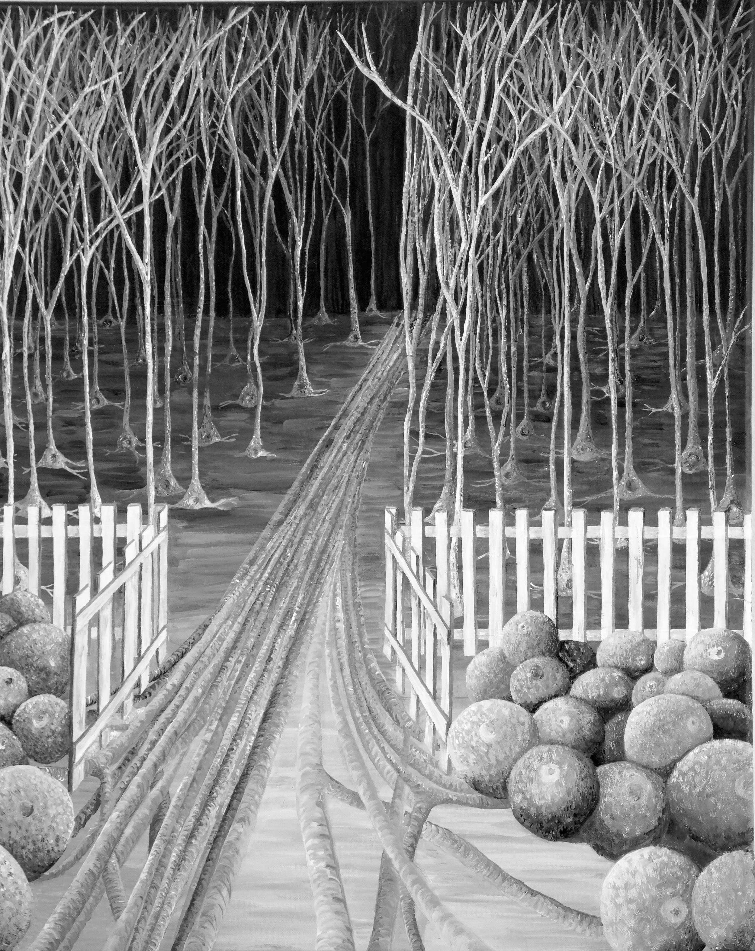
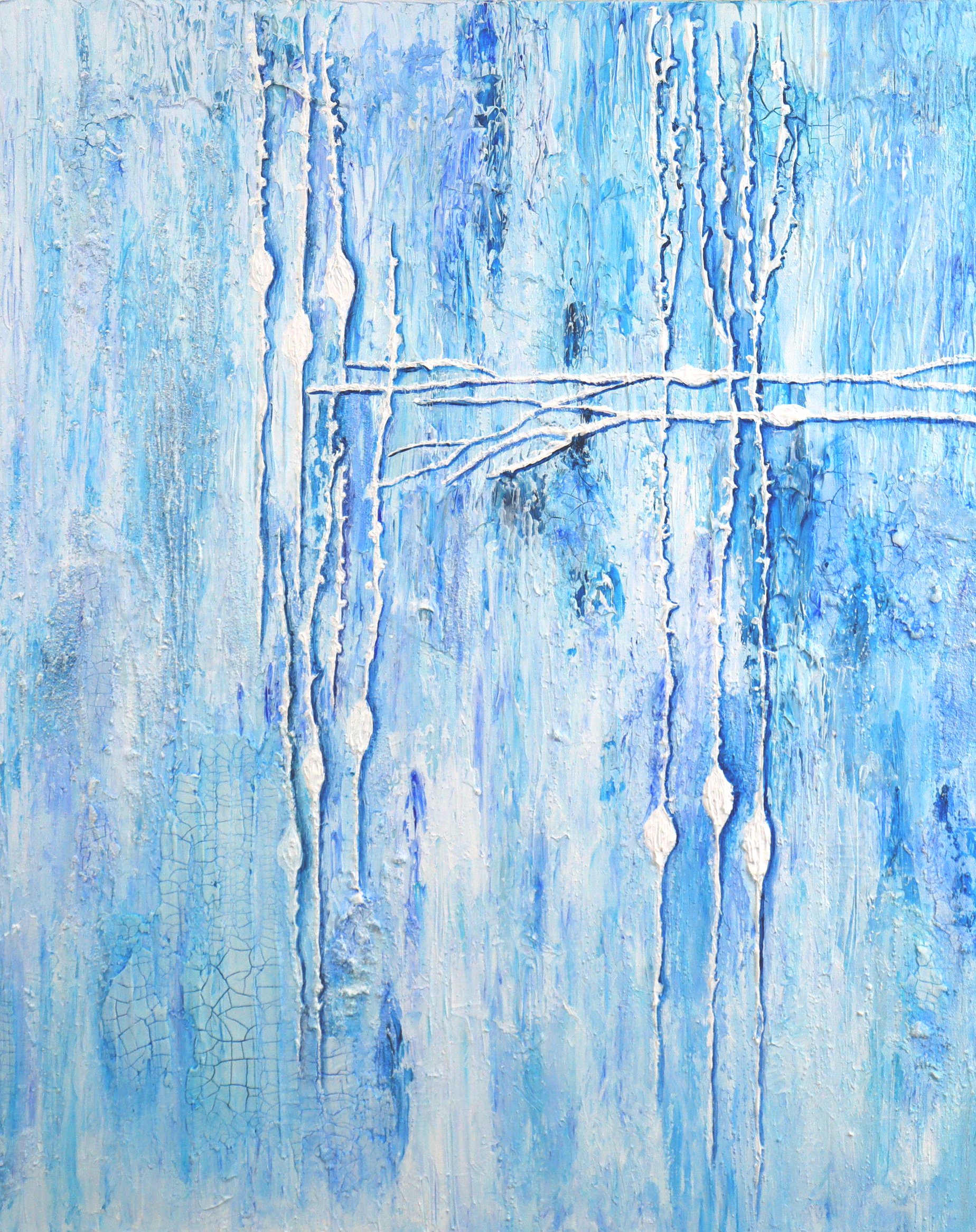
24x30” acrylic painting
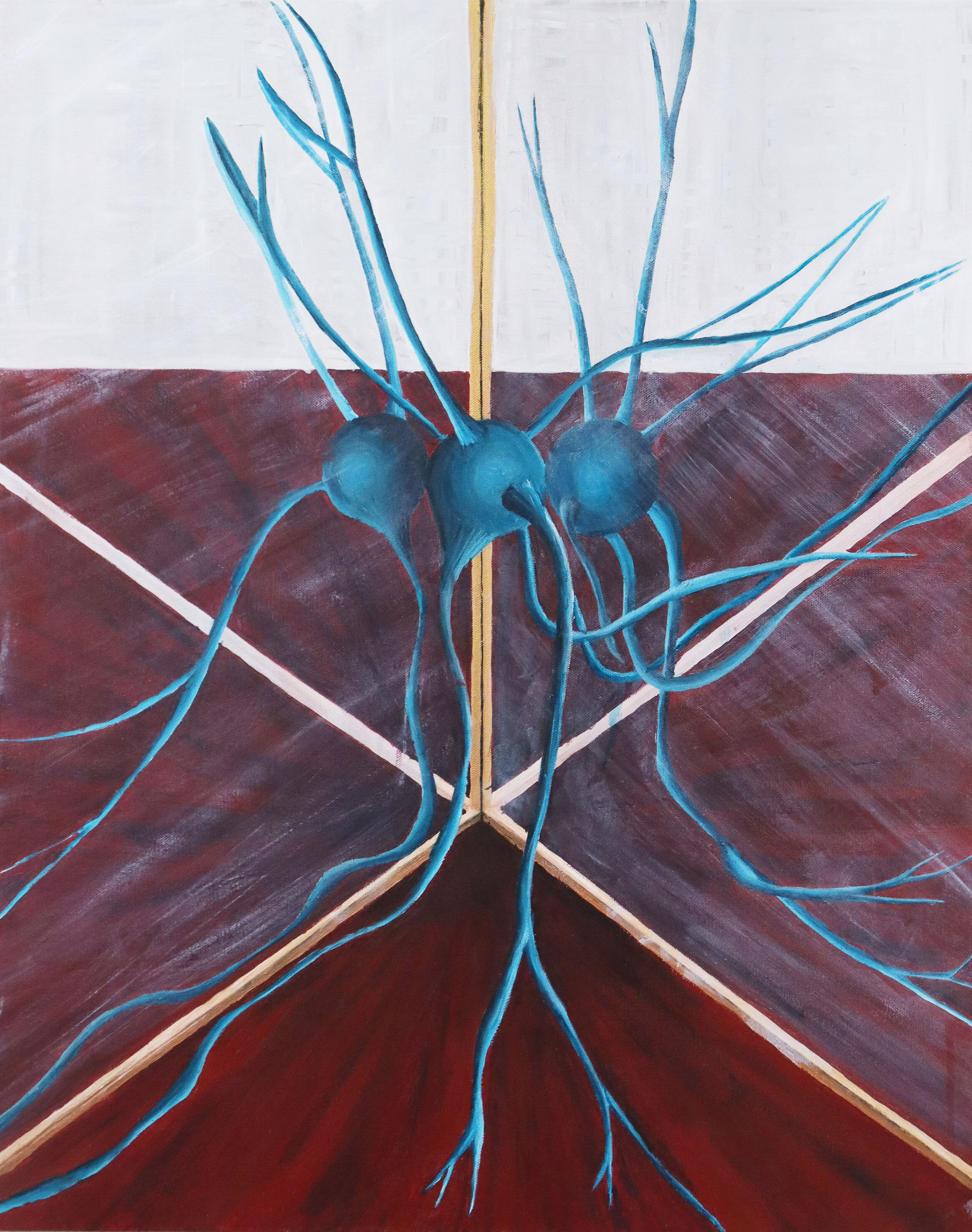
The Gate Control Theory of Pain published in 1965 theorized a balance between excitation and inhibition within the central nervous system. Signals from the sensory neurons (boulders) send signals to the central nervous system (trees) through to modulate pain. The gate can open and close due to activity in different type of neurons.
24x24” acrylic painting on canvas
Neurons
are specialized cells in the nervous system that transmit signals related to nearly all aspects of function: motor, sensation, learning, memory, thoughts, emotion, and much more.
12x36” Acyrlic Painting on Canvas
Dendritic spines, first described by Cajal, are small extensions from the dendrite that make contact with axons. They come in different sizes and shapes.hese spines primarily receive excitatory input. They are plastic and can change shape and volume quickly. Dendritic spines have been implicated in a number of normal processes like learning and memory, as well as disease states like schizophrenia.
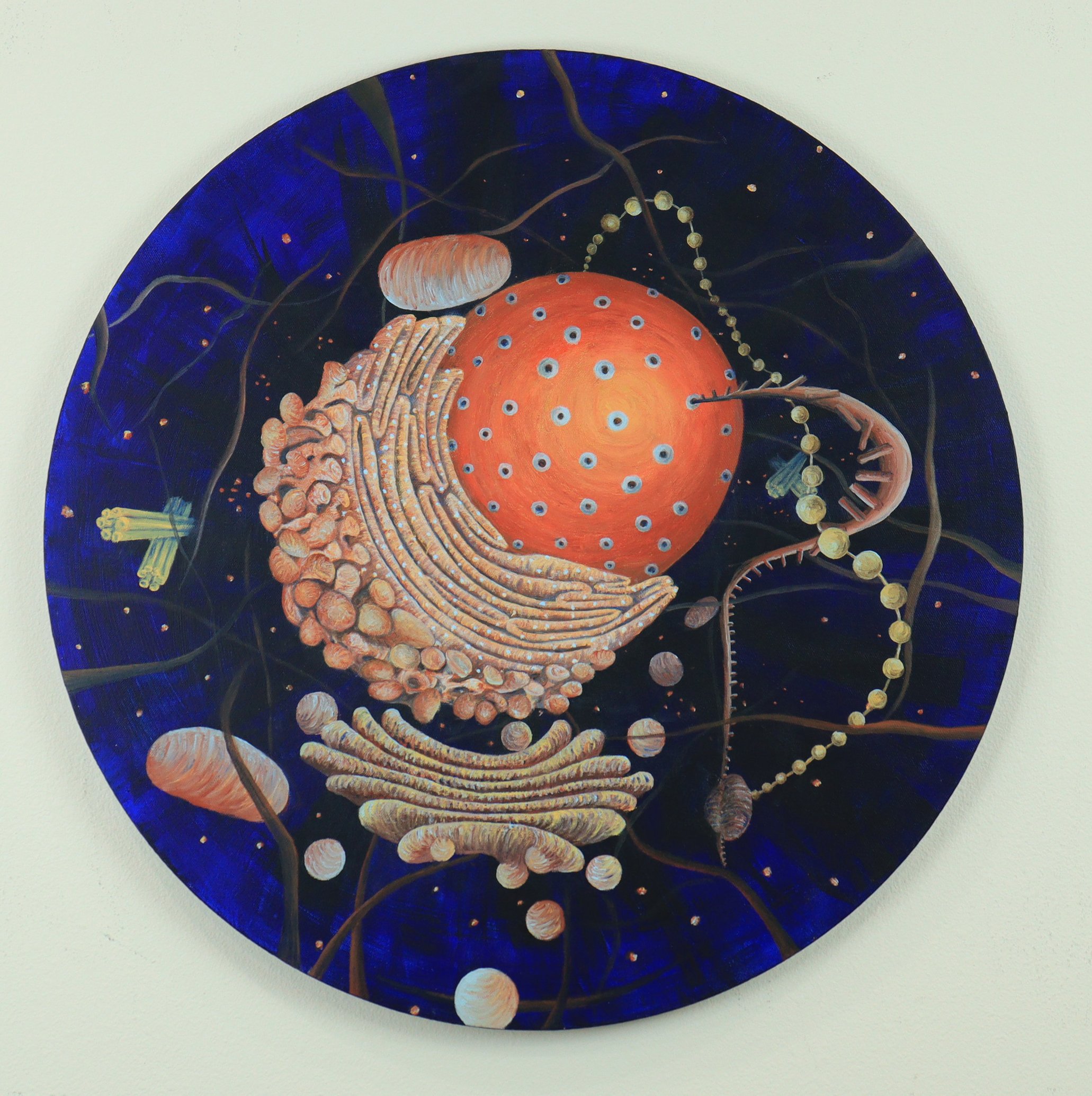
24” round, acrylic on canvas
SOLD
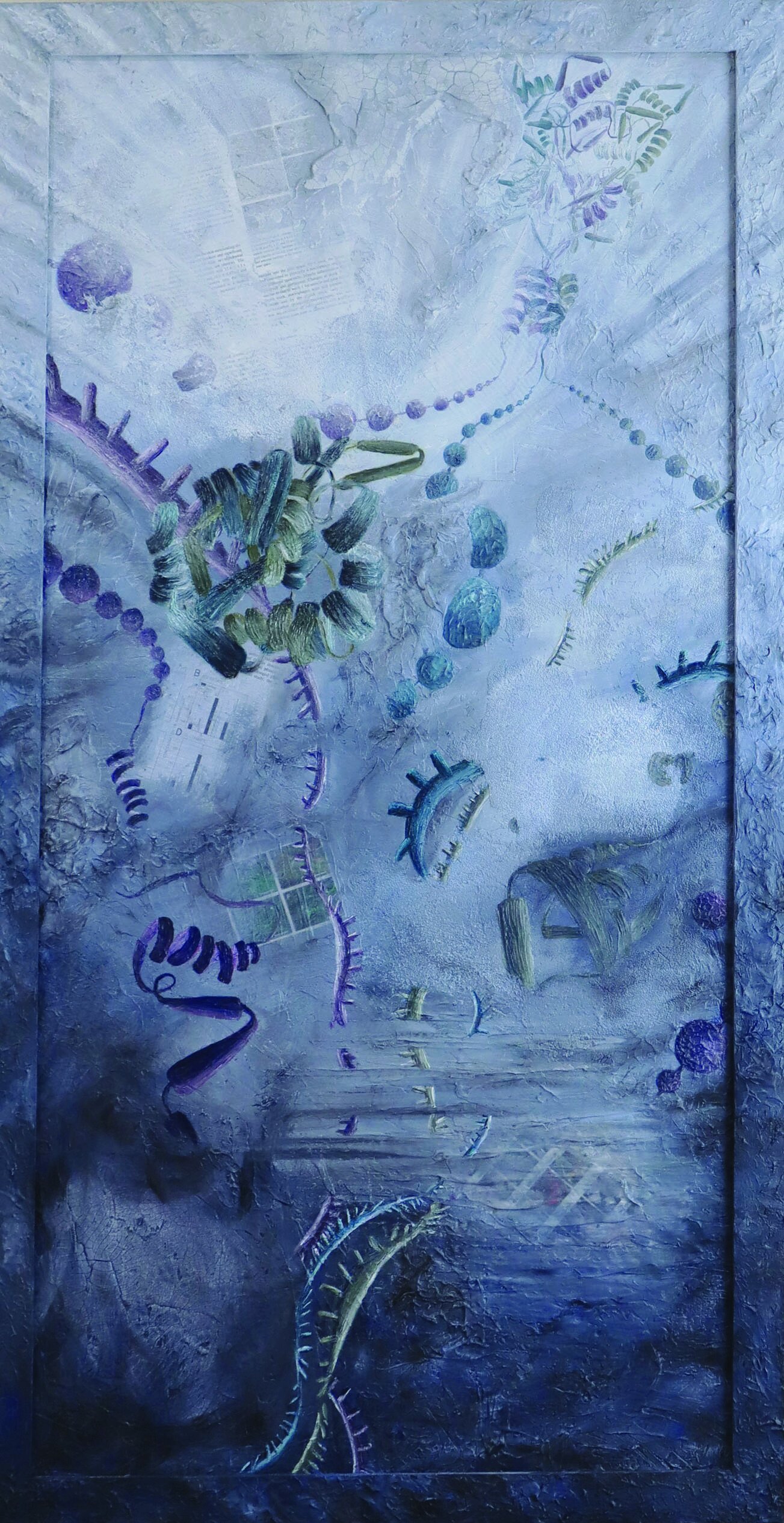
The inside of the cell is an amazing universe of structures and molecules that provide life. The cell, in its nucleus, holds life's code (DNA) and through interaction with several structures converts that code to the proteins that that provide function.
18x24” acrylic painting on canvas
Neurons come in many shapes, sizes, and types. They work together to control movement, sensation, thoughts and memory.
24x30” acrylic on canvas
SOLD
The companion piece to PAIN, Analgesia represents the opposite. While pain is associated with chaos and inflammation, fear, anger and stress, pain relief is associated with order, calm, and peace.
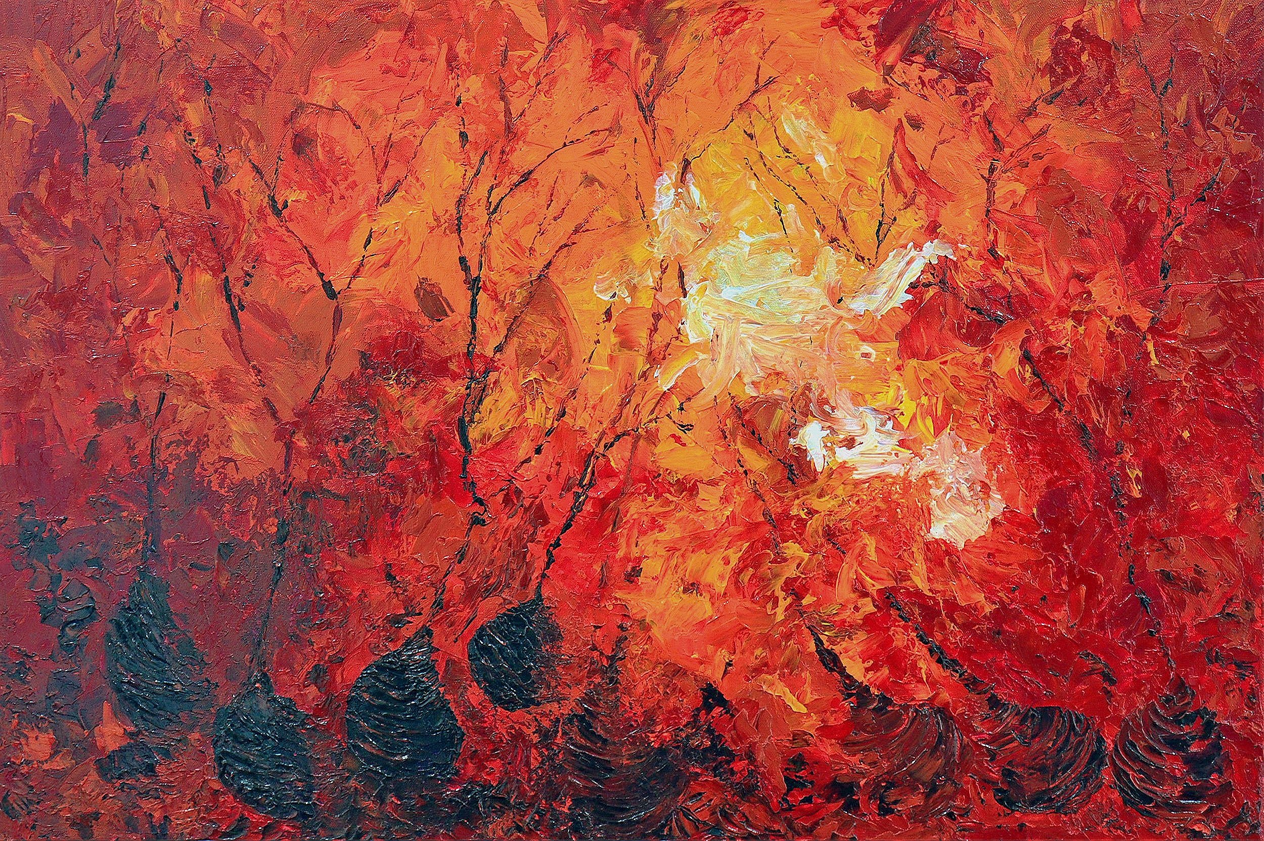
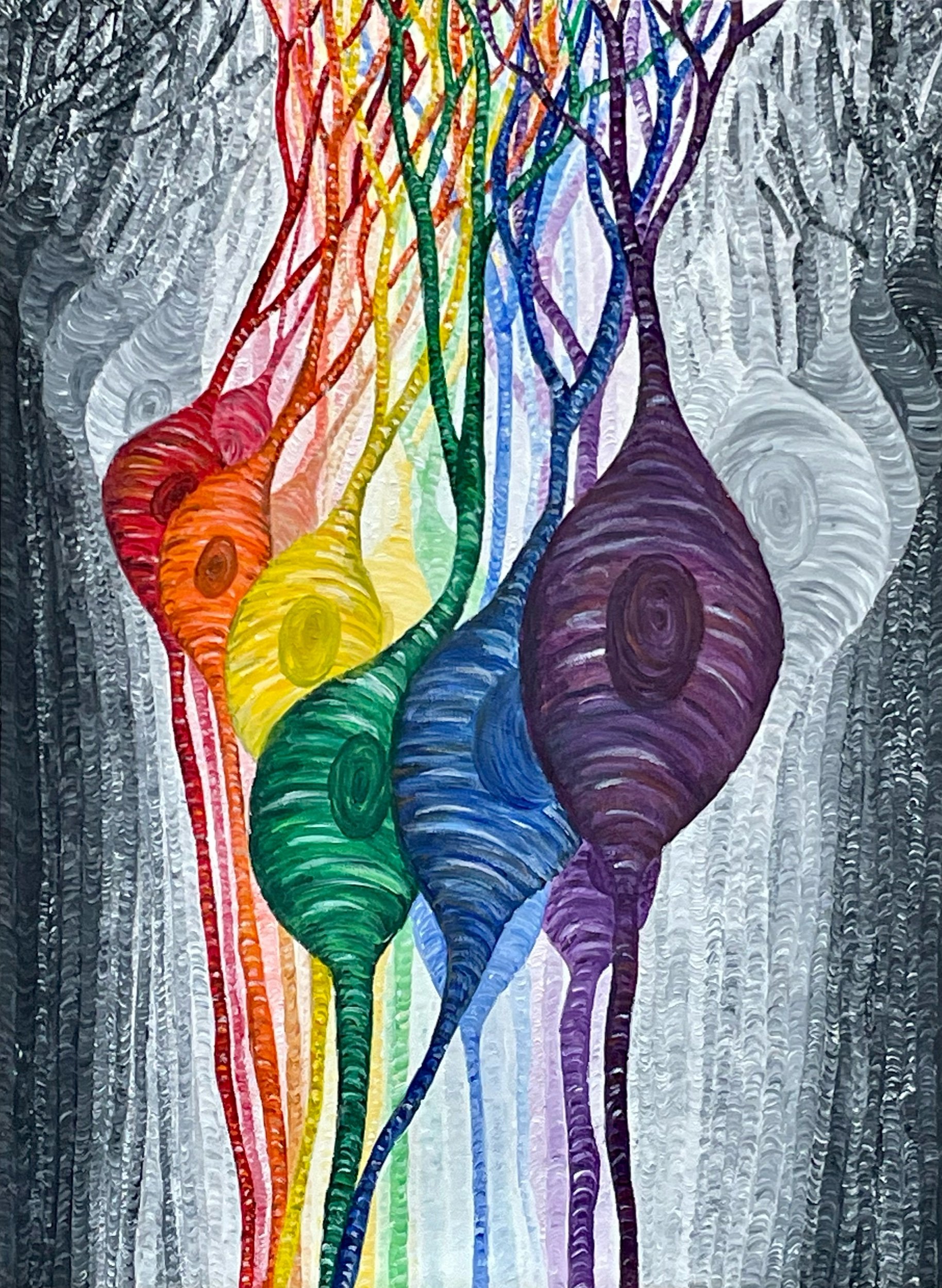
24x36” acrylic painting
SOLD
Chronic pain affects nearly 100 million Americans and costs over 600 billion dollars per year in healthcare and lost wages. Pain is a unique experience that can affect all aspects of a person life from the sensation itself to impacts on work, family and society. Forefront research efforts are under way to develop a better understanding of pain and novel approaches to its management.
16x20” metallic acrylic painting on canvas
SOLD
Muscle spindles are located within every muscle and are responsible for your ability to sense your limb in space. They are found within an independent capsule and run parallel to the main muscle fibers. There are two types nuclear bag and nuclear chain that respond to stretch and length of changes in muscle.
SOLD

9x12” multimedia
SOLD
Neurons carry signals from one area to the next by axons and receive information through their dendrites. The background is a collage of published manuscripts, glazed with turquoise paint, that examine neuron function. Neurons were painted with acrylics over the top of the manuscripts.
39”x55” multimedia (collage/acrylic)
SOLD
Immune cells fight infection and injury, maintain homeostasis and are connected to all parts of the body through the blood stream. There are a variety of immune cells that each confer a different action to respond to infection and injury. Many chronic conditions have alterations in the immune system. There is extensive research into the study of the immune system in disease, including chronic pain.
SOLD
24x48” multimedia
Proteins are large molecules that are critical to the structure, function and regulation of the body’s tissue and organs. Protein synthesis occurs in two stages, transcription and translation. Transcription refers to the process where the genetic instruction found in the DNA is transferred to messenger RNA. Translation is the process where messenger RNA codes for amino acids that form a chain or polypeptide that subsequently combine together to form a protein molecule.
8x10” acrylic painting on clayboard
SOLD
Neurons make intricate connections to other neurons through their dendrites.
Acrylic on Wood.
The immune system is involved in normal response to injury and healing. Increasingly, alterations in the immune system have been found to play a role in many diseases. Cytokines, released from immune cells, are proteins responsible for the actions of the immune system. They can respond in an exaggerated way, referred to as a cytokines storm, making diseases worse.

24x36” acrylic painting
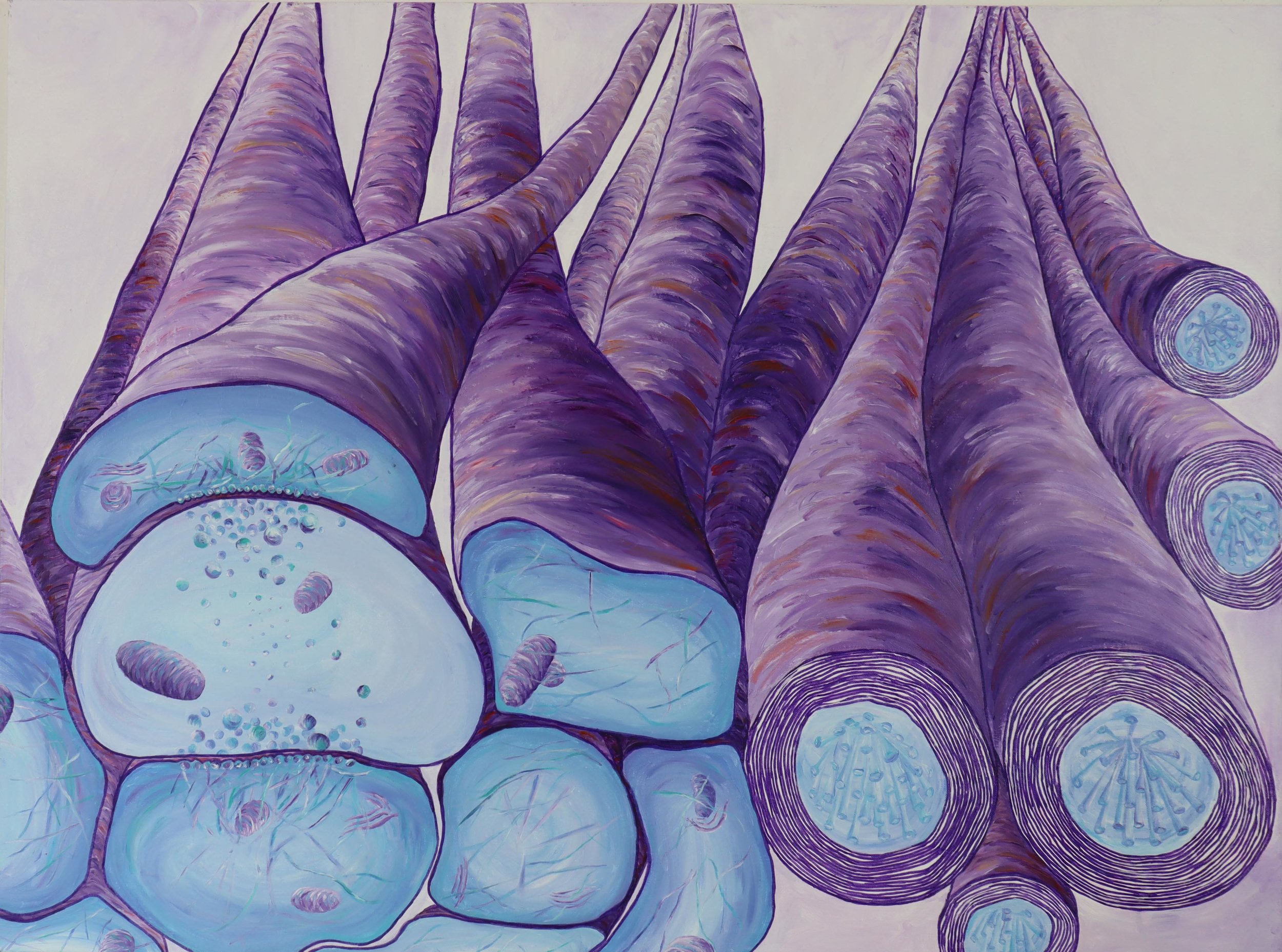
Skeletal muscle cells are incredibly large with diameters of up to 100 micrometers and up to 30 cm long. Muscle fibers are arranged in bundles, called fascicles, and multiple fascicles form muscles that provide the means for us to move our joints. Skeletal muscle appears striped because of the regular arrangement of proteins involved in muscle contraction, actin and myosin.
16x20” acrylic painting
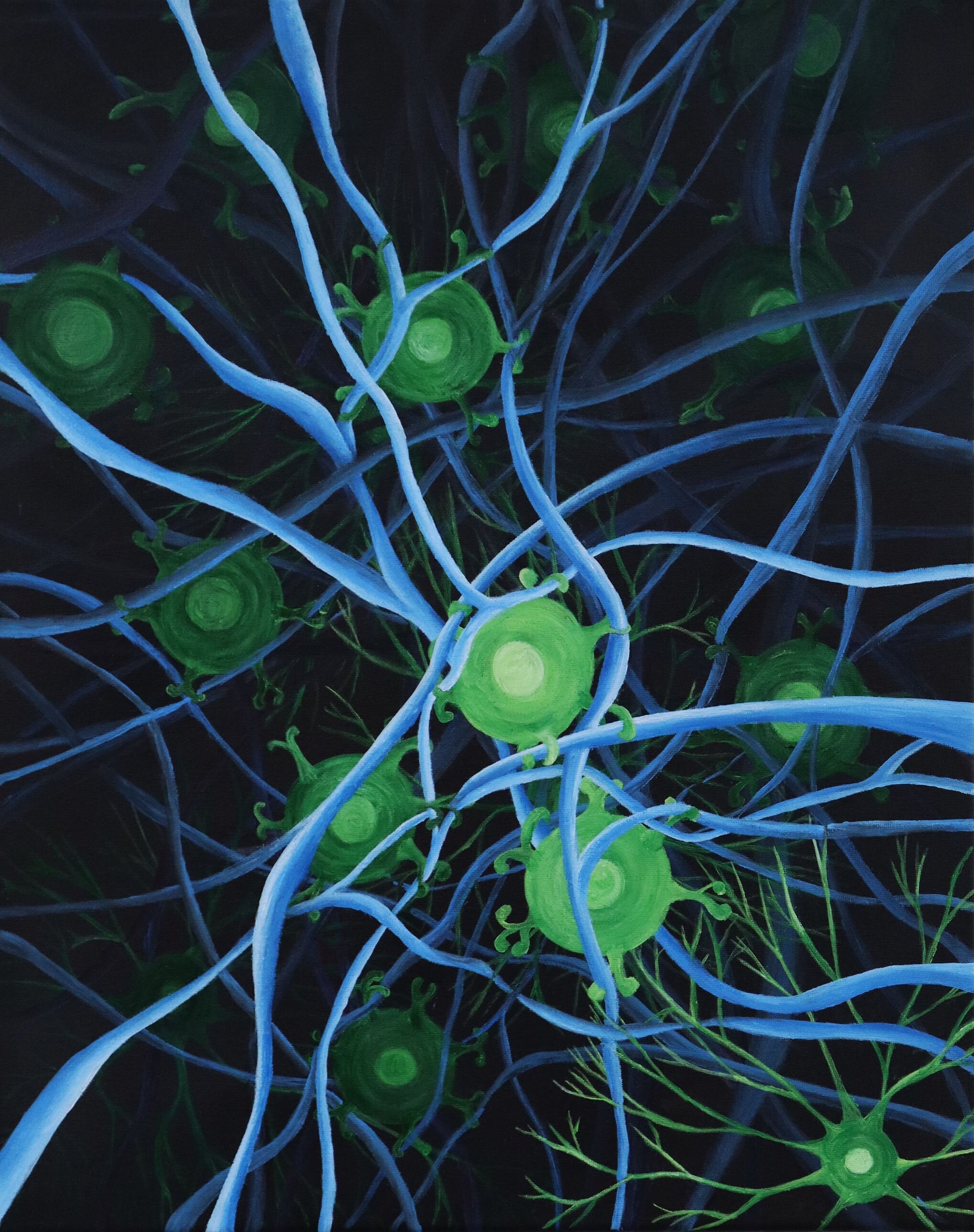
Microglia in the brain play a significant role in many nervous system diseases including pain and depression. When activated their cells bodies become larger and the processes retract. They communicate with neurons and their axons through release of chemicals. Targeting microglia in the future may be able to treat a number of nervous system diseases.
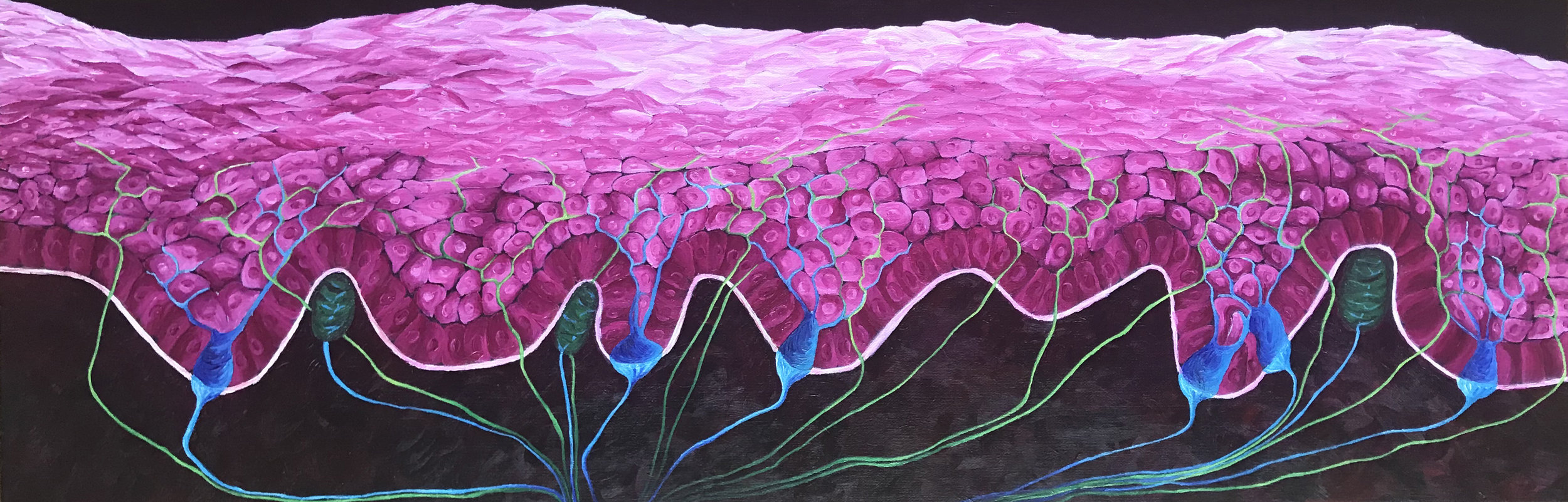
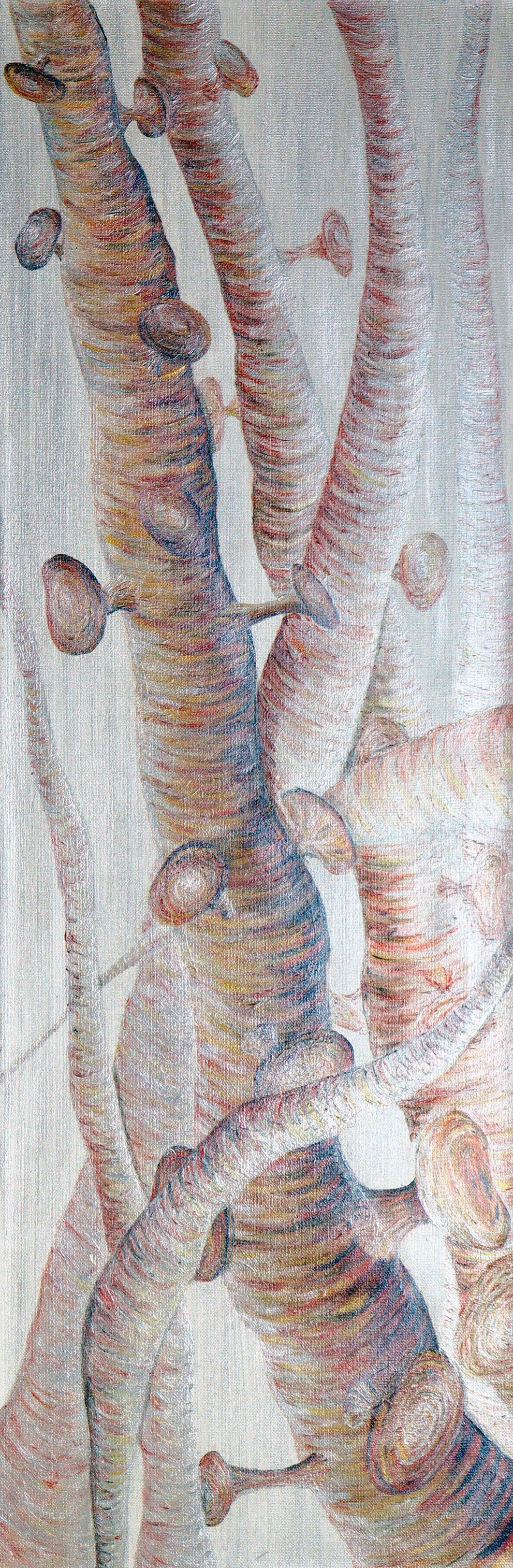
12x36” Acrylic Painting
The epidermis, in pink, is separated from the dermis, in black, by the basement membrane in white. The epidermis is constantly making new cells from its basal layer, the dark pink column-like cells on top of the white basement membrane. Pain nerves, in green, course through the epidermis. Meissner’s corpuscles, dark green ovals in the dermis, are responsible for light touch. They are very sensitive and found in the highest concentrations in your fingers and lips.
30x24" acrylic painting
SOLD
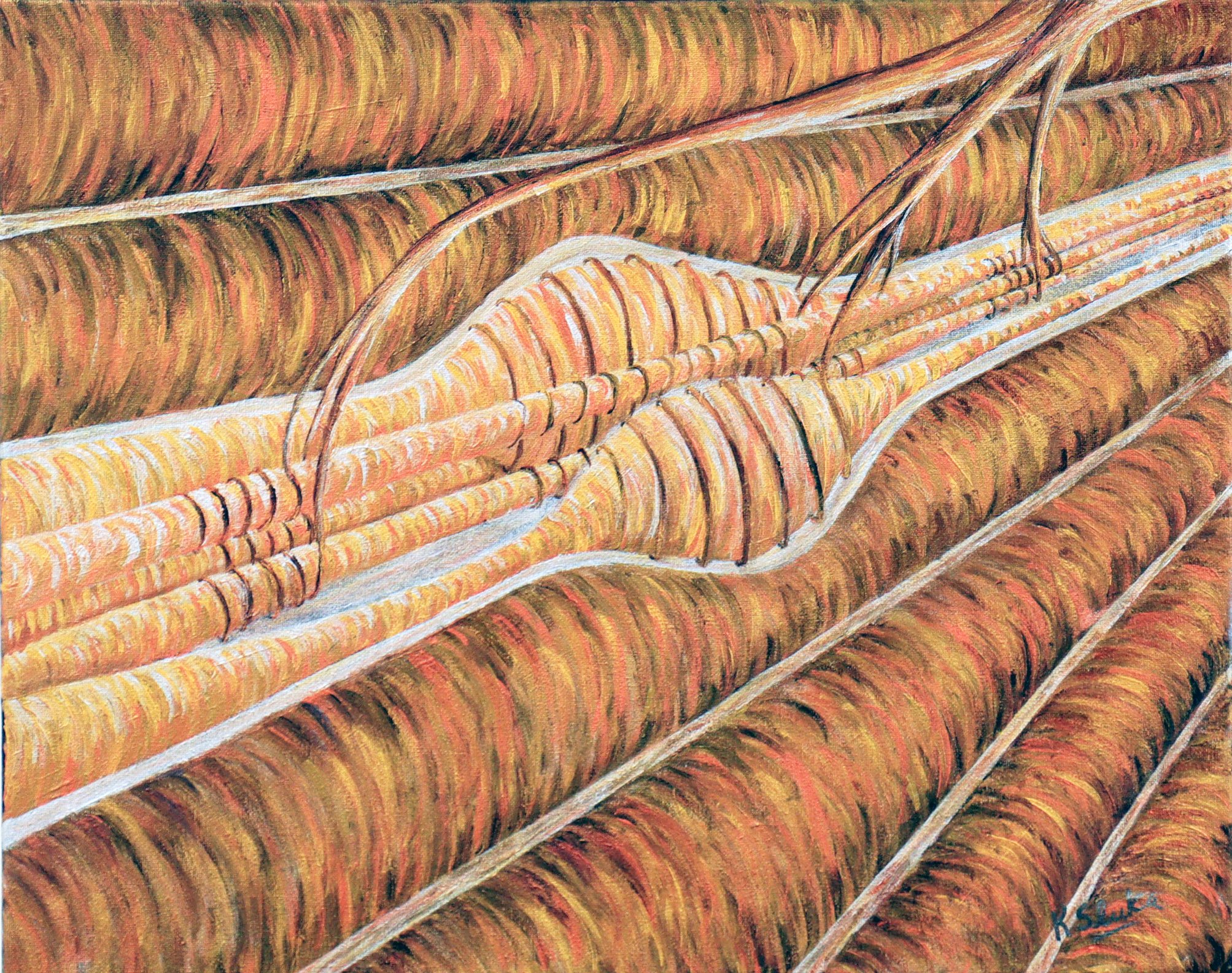
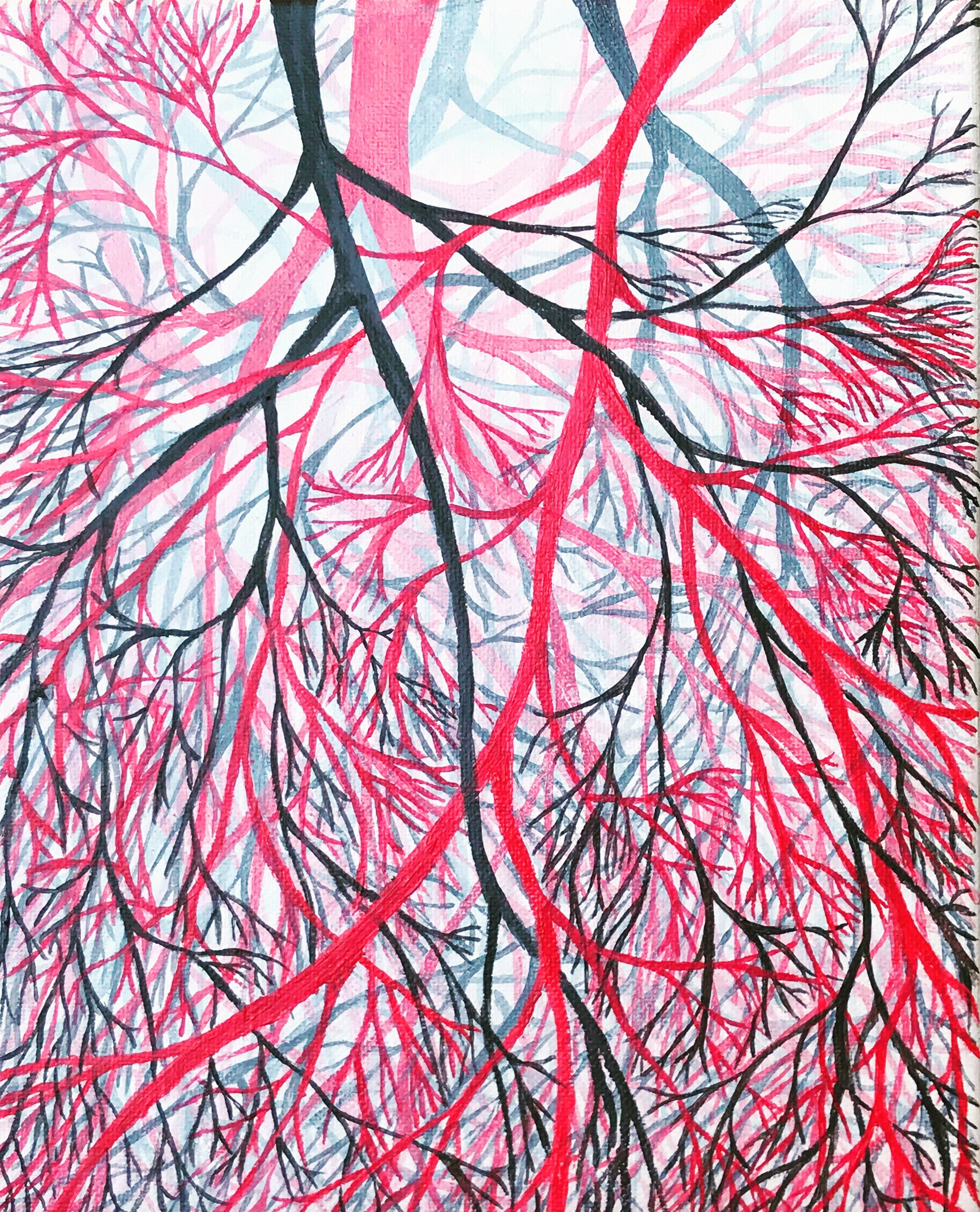
Peripheral nerves branch into smaller divisions as they dive off to innervate our tissue. They send branches to muscles to promote movement, and receive input from skin, muscles, joints, connective tissue, and bone for us to interpret the environment around us. Sensations of touch, pressure, pain, and more are carried through these axons to their cells bodies in the dorsal root ganglia-background of the painting, and then on to the central nervous system.
30x40” Acrylic Painting
Neural circuits are population of neurons that are connected by synapses to carry out specific functions. The brain functions as a very sophisticated computer that can modify signals through excitatory and inhibitory neural circuits. Neural circuits connect brain areas to result in learning, thoughts, emotions, movements, and sensations.
18x24” acrylic painting
SOLD
Heat maps are used in large data sets like proteomics or transcriptomics. These represent genes or proteins that are upregulated or downregulated. These platforms are great for discovering novel mechanisms of disease particularly between male and female sexes.
SOLD
16x20” acrylic painting
Stem cells give rise to a number of different cells types. Stem cells can differentiate into endothelial cells that line blood vessels, neurons, epithelial cells that line the gut, adipocytes (fat), muscle cells, cartilage or immune cells. Tissue specific signals provide information to stem cells for them to become specialized for a particular function.
24x30” Acrylic Painting
Mirror neurons are brain cells that are activated when a particular action is performed and when that action is observed in others. Mirror neurons are proposed to be the neural basis for empathy.
18x24” Acrylic Painting
Neurons form complex networks for communication. Each neuron may have as many as 15,000 connections with other neurons. Electrical signals, action potentials, are transmitted along the axon to a synapse. When the electrical signal reaches a synapse it releases chemical signals called neurotransmitters. The neurotransmitter diffuses across the synapse and binds to receptors on another neuron to transmit the messsage.
24x28" Acrylic Painting
SOLD
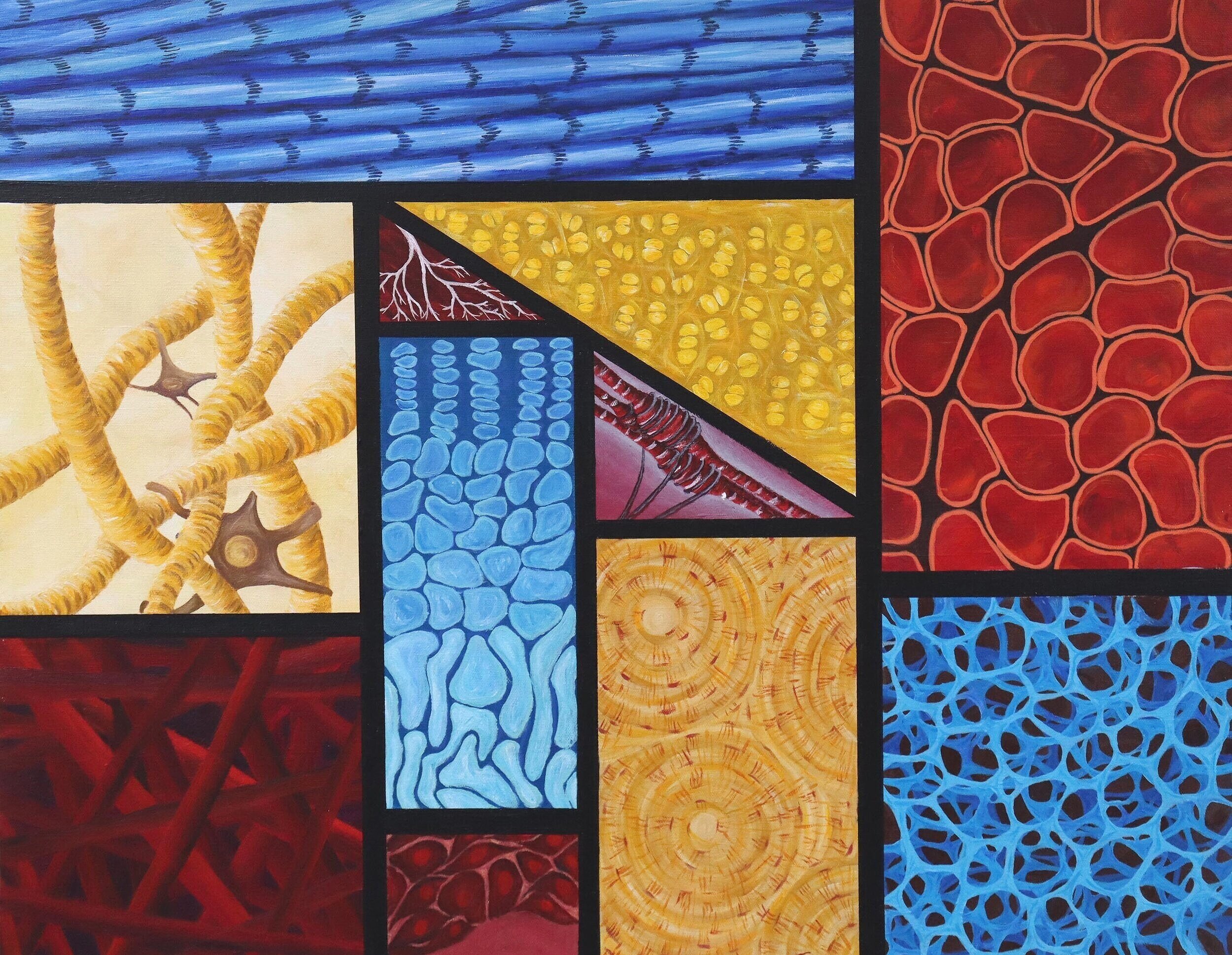
The musculoskeletal system is composed a variety of tissue types designed to promote movement. This painting is designed based on Piet Mondrian’s style using primary colors and geometric shapes. Within each square I show a tissue or cell type related to the musculoskeletal system in primary colors. These include, tendon, muscle, bone, connective tissue, synovium, nerve, cartilage, and muscle tissue.
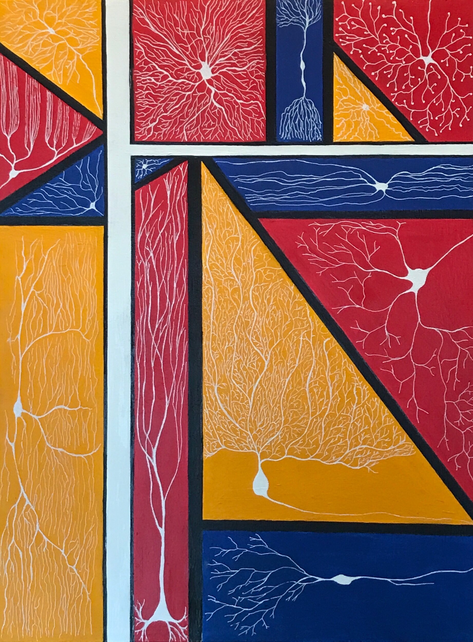
18x24” Acrylic painting
SOLD
An abstract design fashioned from work of Piet Mondrian.
Inside each shape is a different type of neurons. Neurons come in many sizes and shapes which are likely important for their function.
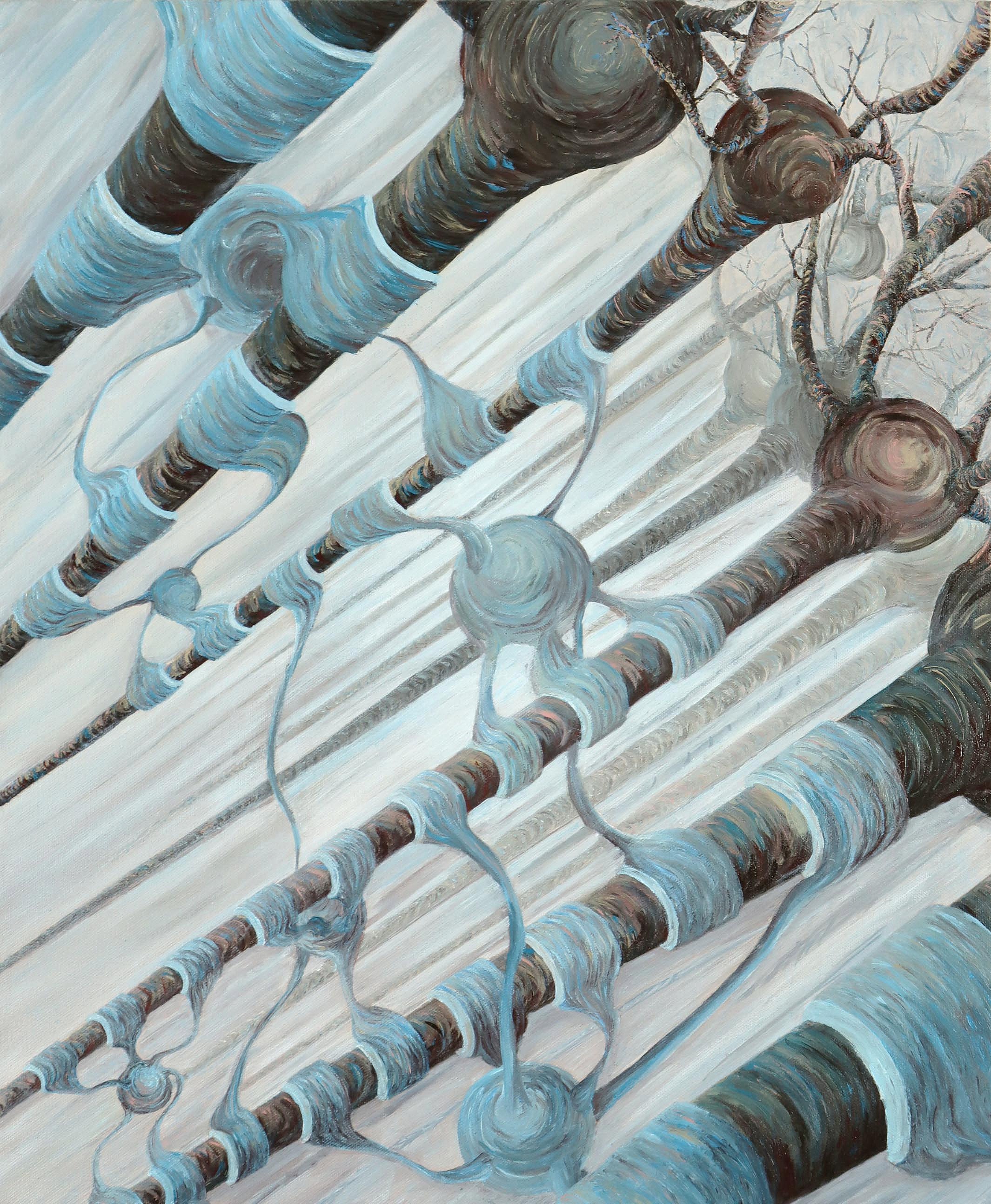
20x24” Acrylic Painting
Neurons send electrical signals through their axons, in brown. The speed of the electrical signal is determined by the myelin sheath which is made from a glial cell called an oligodendrocytes (blue). The myelin is considered insulation and speeds the conduction of electrical signals down the axons.
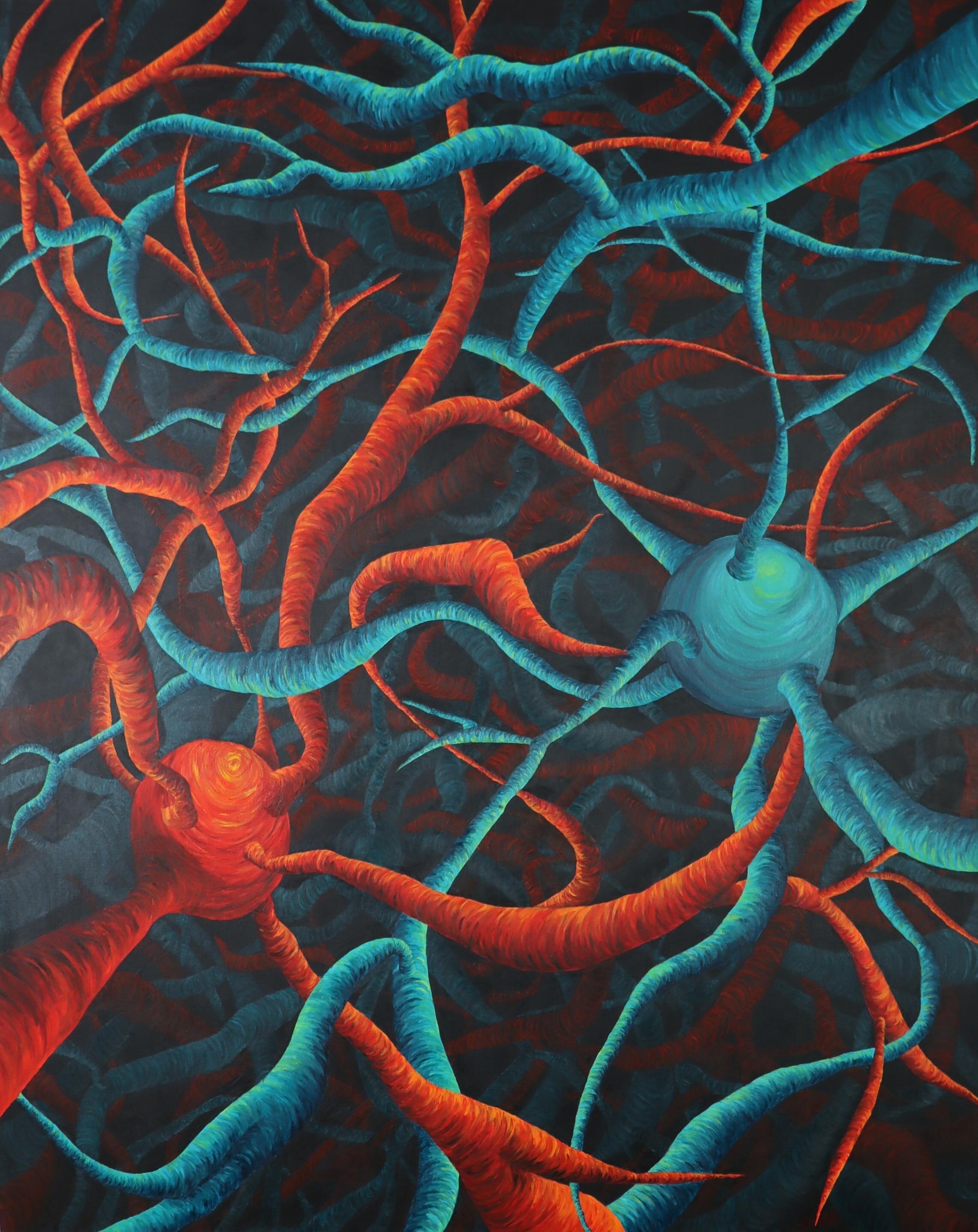
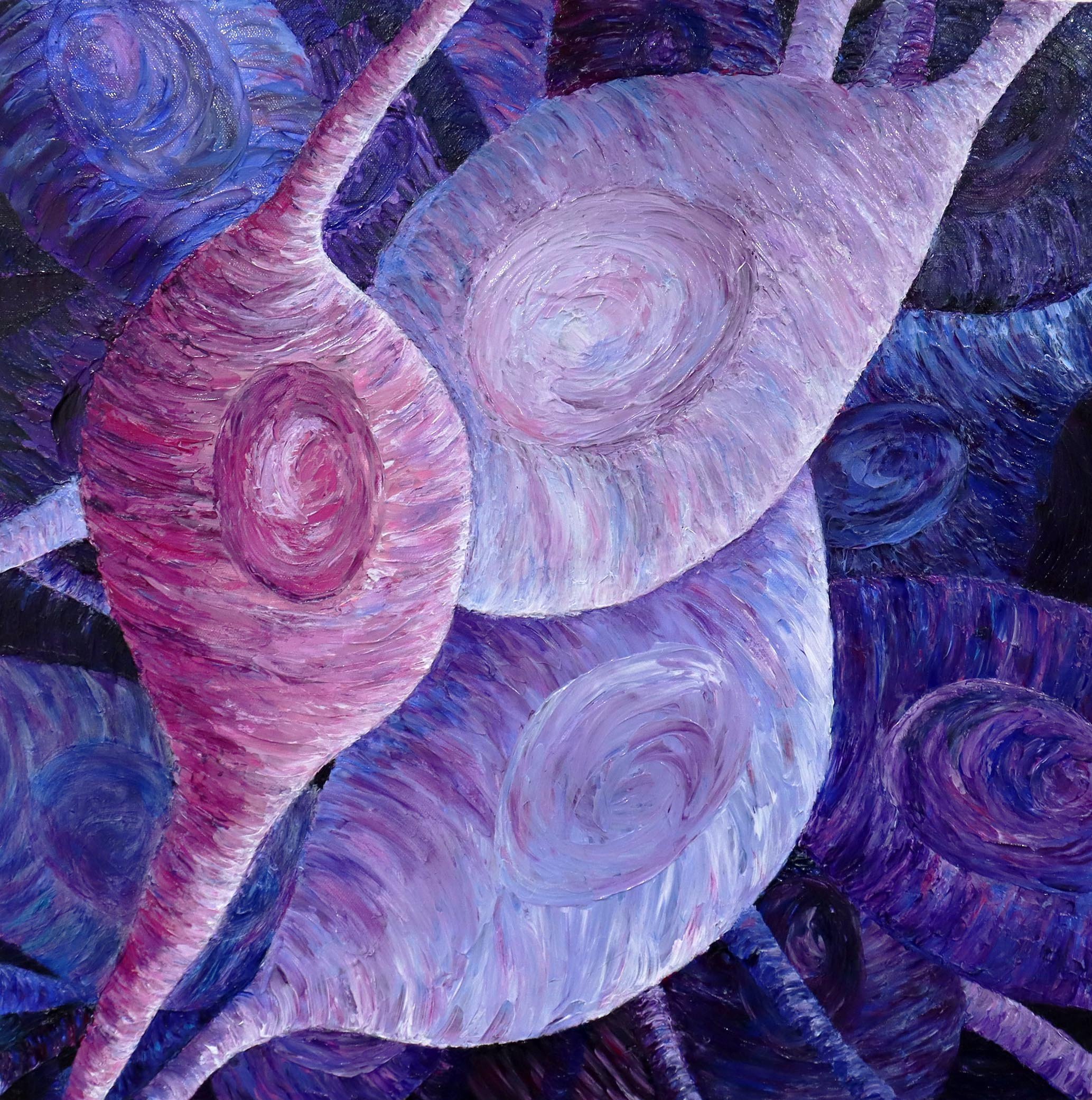
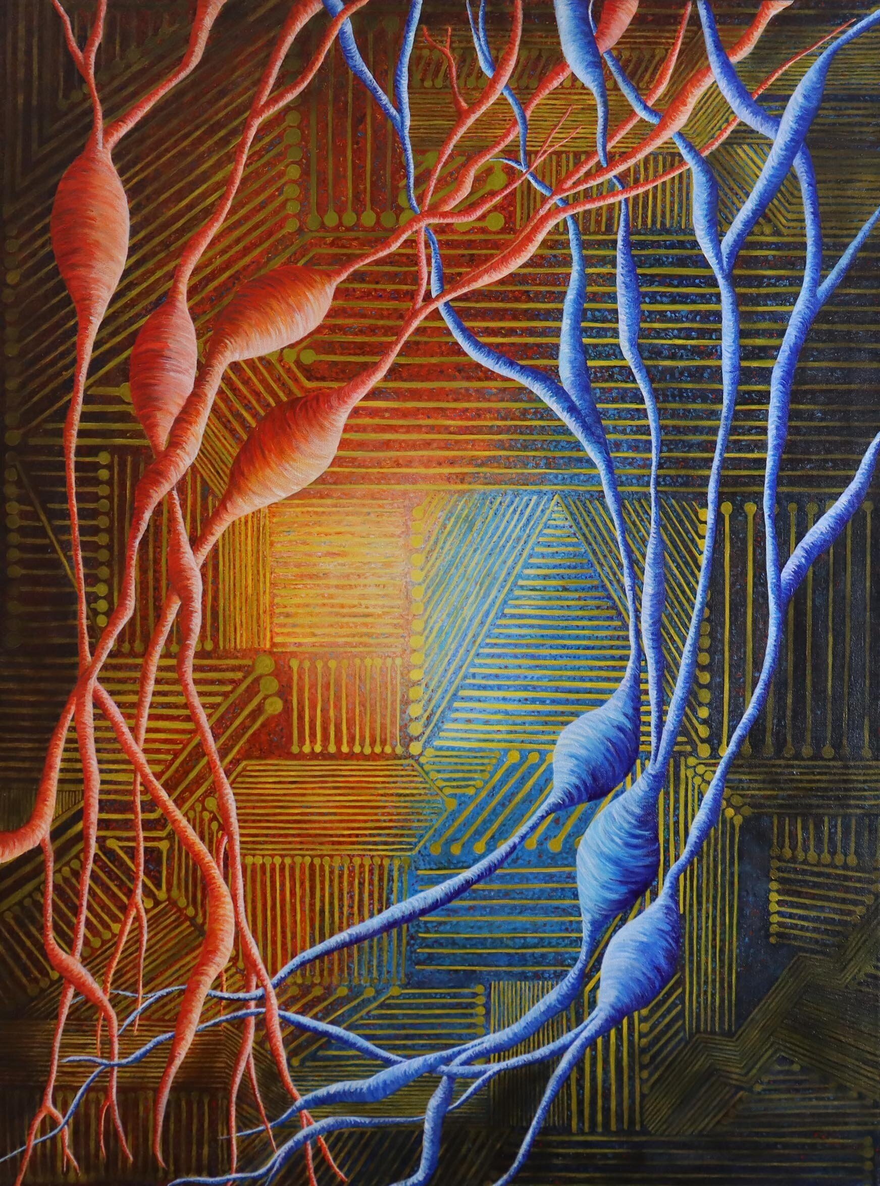
4x5’ Acrylic Painting
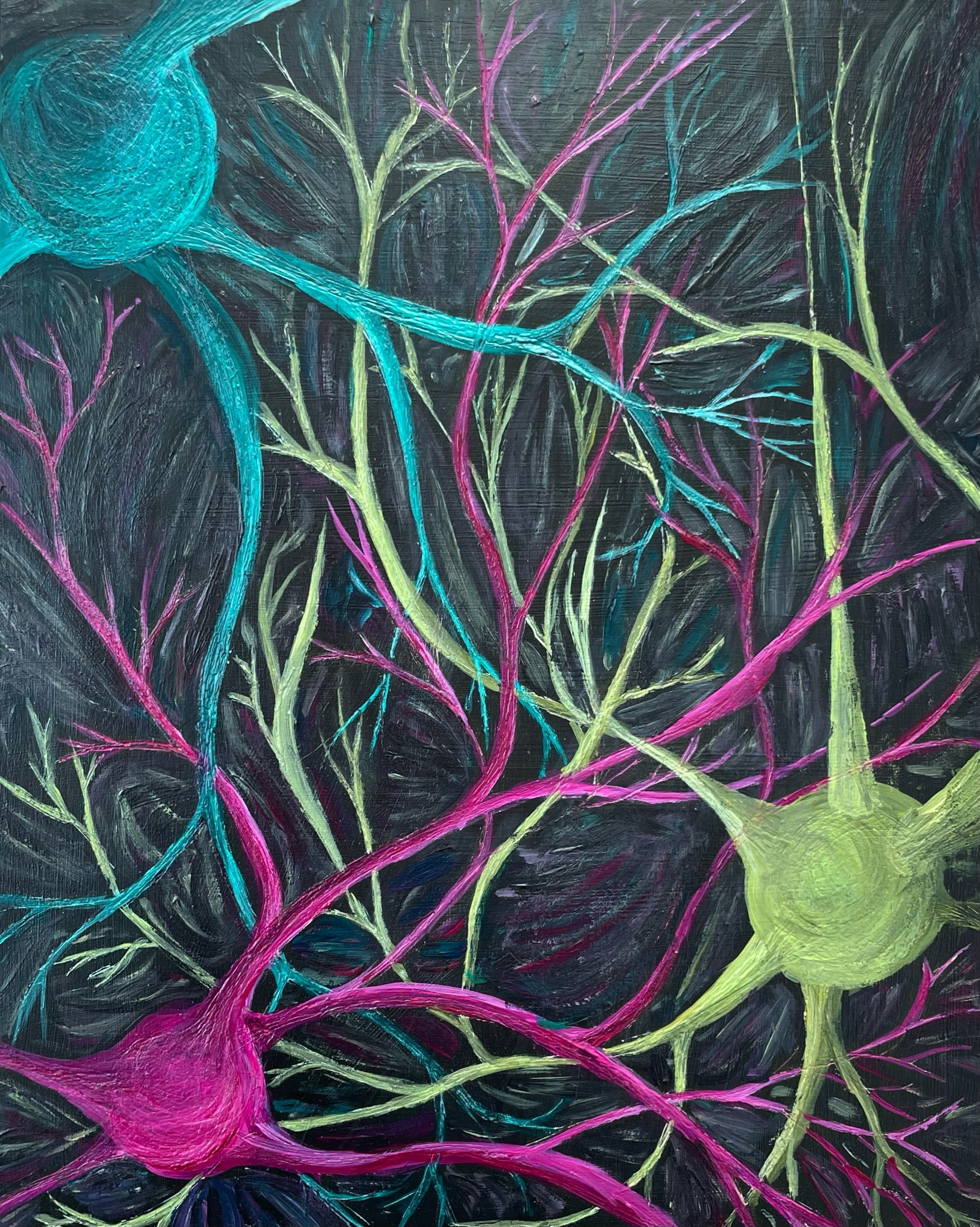
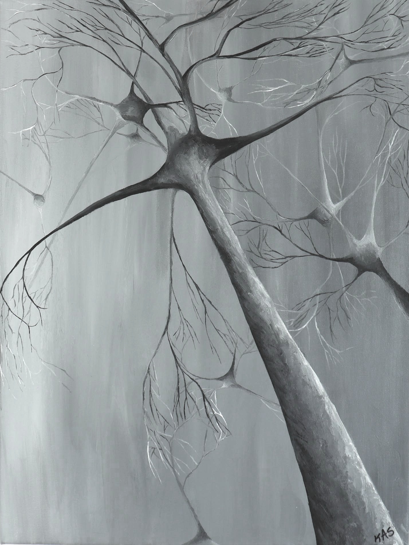
Neurons communicate with other neurons through their axons and dendrites. The dendrites extend from the round cell body and branch like trees to form a tangled web of connections with other neurons. This painting highlights two neurons, in red and turquoise. Additional neurons and dendrites in the background illustrate the depth of field of the neuromatrix and show the complicated web of dendrites that give rise to all that our brain does.

16x20” Acrylic Painting
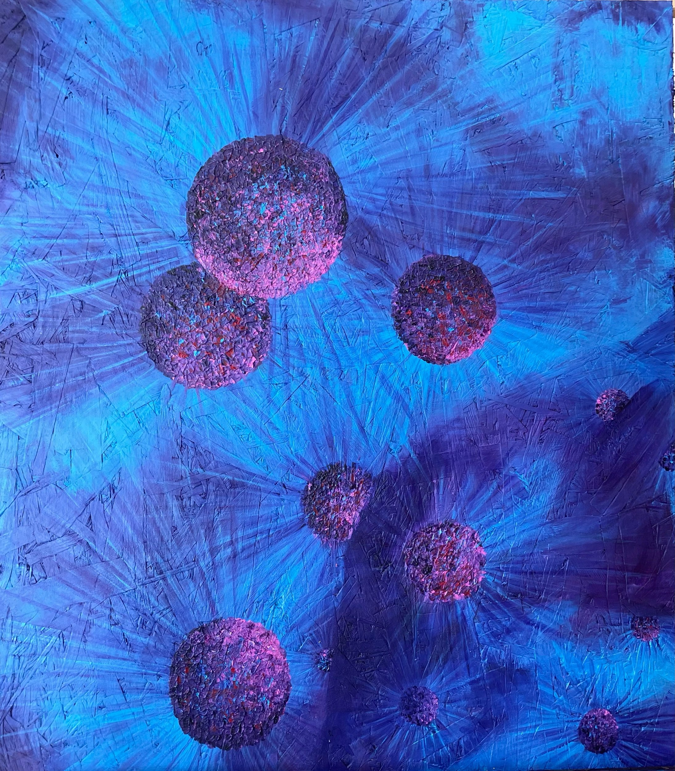
Cancer involves abnormal cell growth with the potential to invade or spread to other parts of the body. Cancer is a group of diseases with over 100 different types.
Treatment for many forms of cancer has improved exponentially over the last few decades, greatly decreasing the number of deaths caused by cancer. Because cancer is a group of diseases it is unlikely there will ever be a single “cure”.
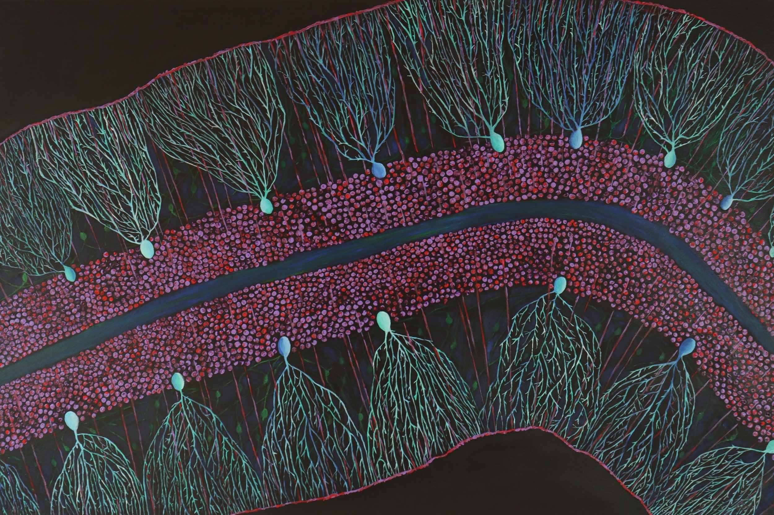
24x36” Acrylic Painting
SOLD
The cerebellum, which is involved in coordination of movement, has a very organized structure that includes the large Purkinje cells (blue/green cells), granular cells (pink/purple cells), and basket cells (small green cells).
Purkinje cells (blue/green cells) receive more synaptic inputs than any other type of cell in the brain—with a single cell receiving more than 200,000 inputs. The granule cell (red/purple) on the other hand is one of the smallest and most numerous cells in the brain.
SOLD
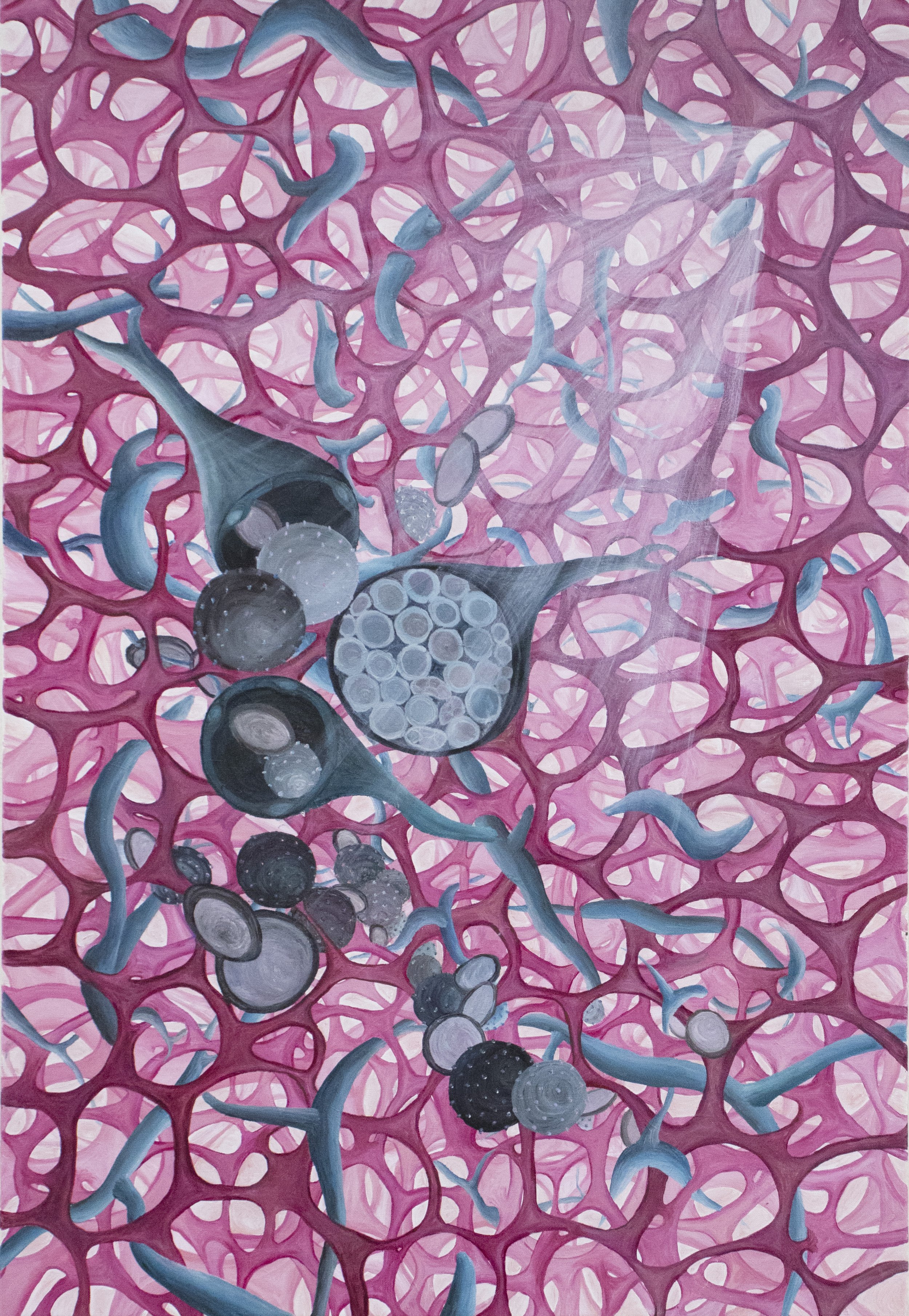
30x40” Acrylic Painting
Neurotransmission is the process of communication between neurons. Axons from one neuron connect to dendrites from other neurons to relay signals through a synapse. One side of the synapse contains the chemicals, called neurotransmitters, in round vesicles. Mitochondria, oval shaped, provide energy for the process. Actin, squiggly lines in dendrites, provides support and structure. On the right are axons with the myelin sheath, round circles, which speed the electrical signal.
16x20” Acrylic Painting
SOLD
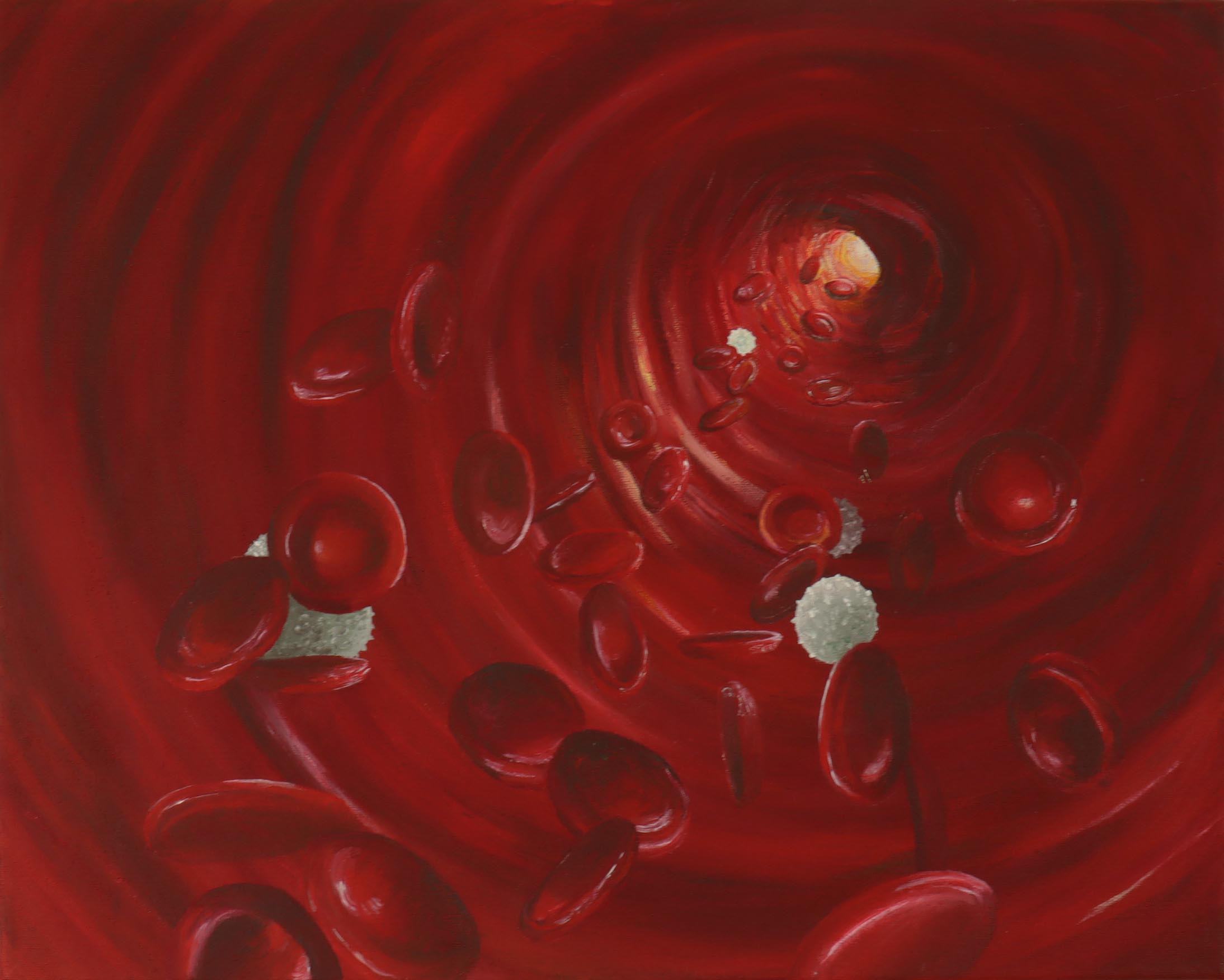
This acrylic painting shows the inside of a blood vessel. Red blood cells carry hemoglobin throughout the body. White blood cells are immune cells that protect the body from infection.
18x24” Acrylic Painting
SOLD
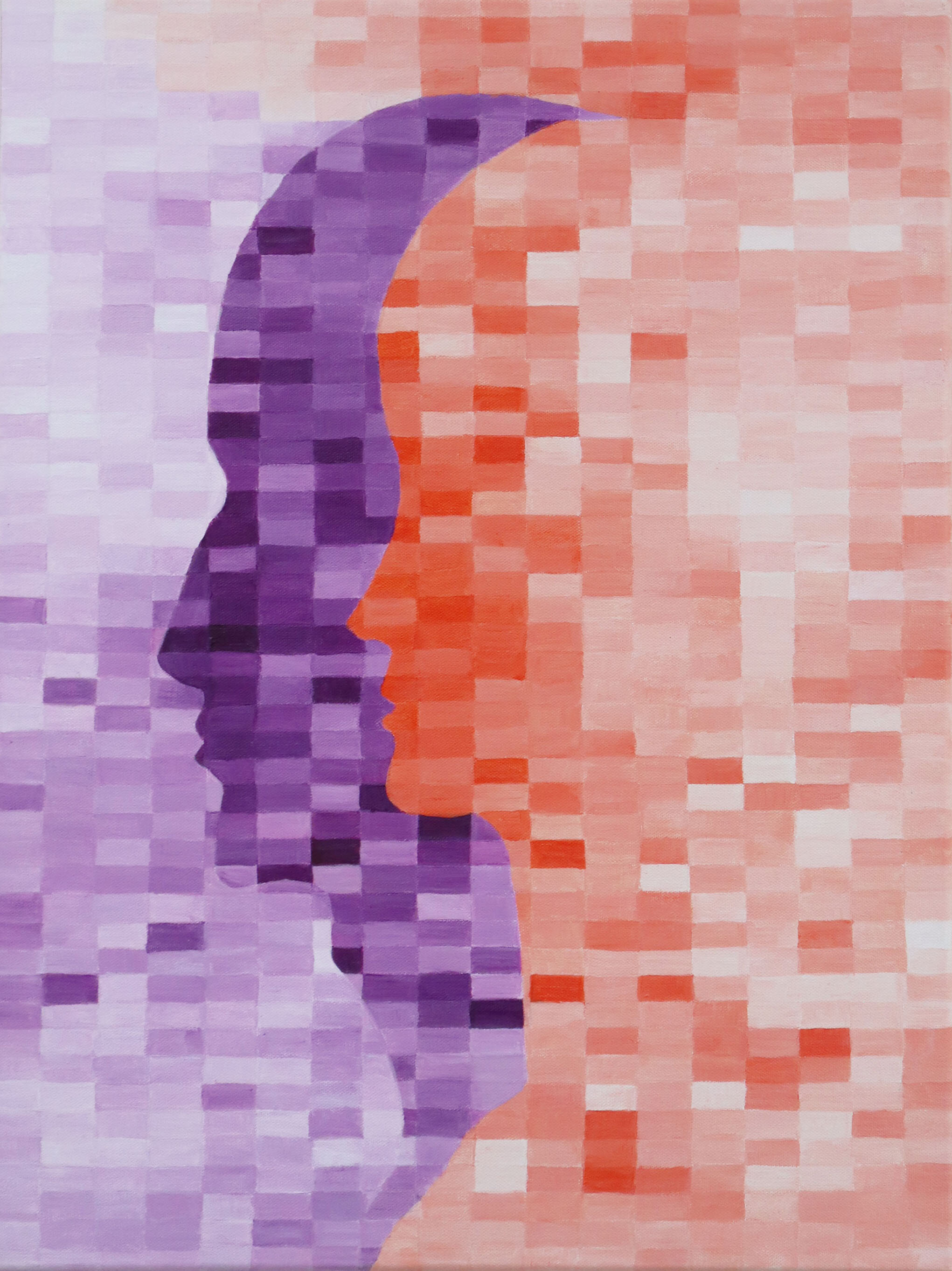
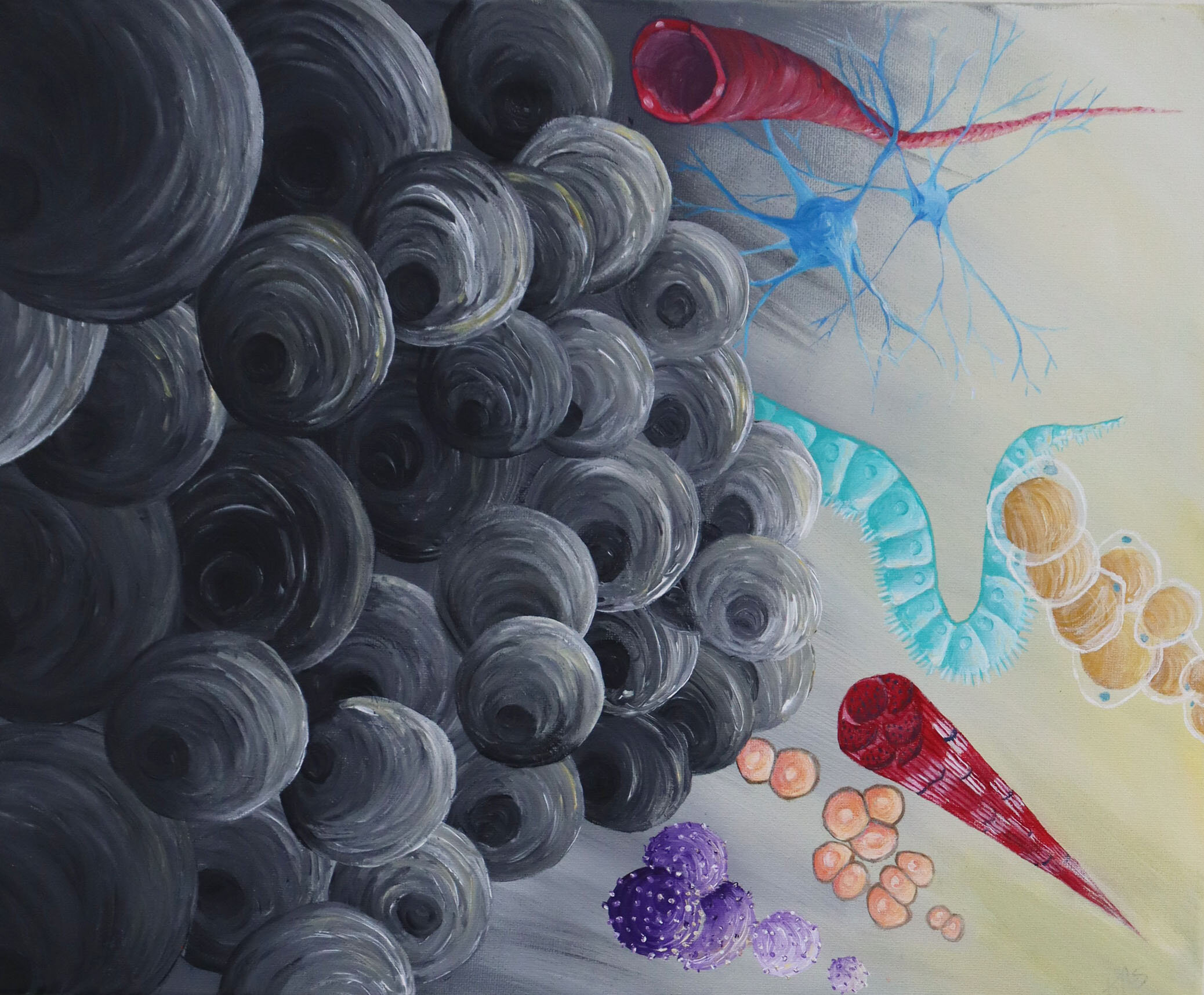
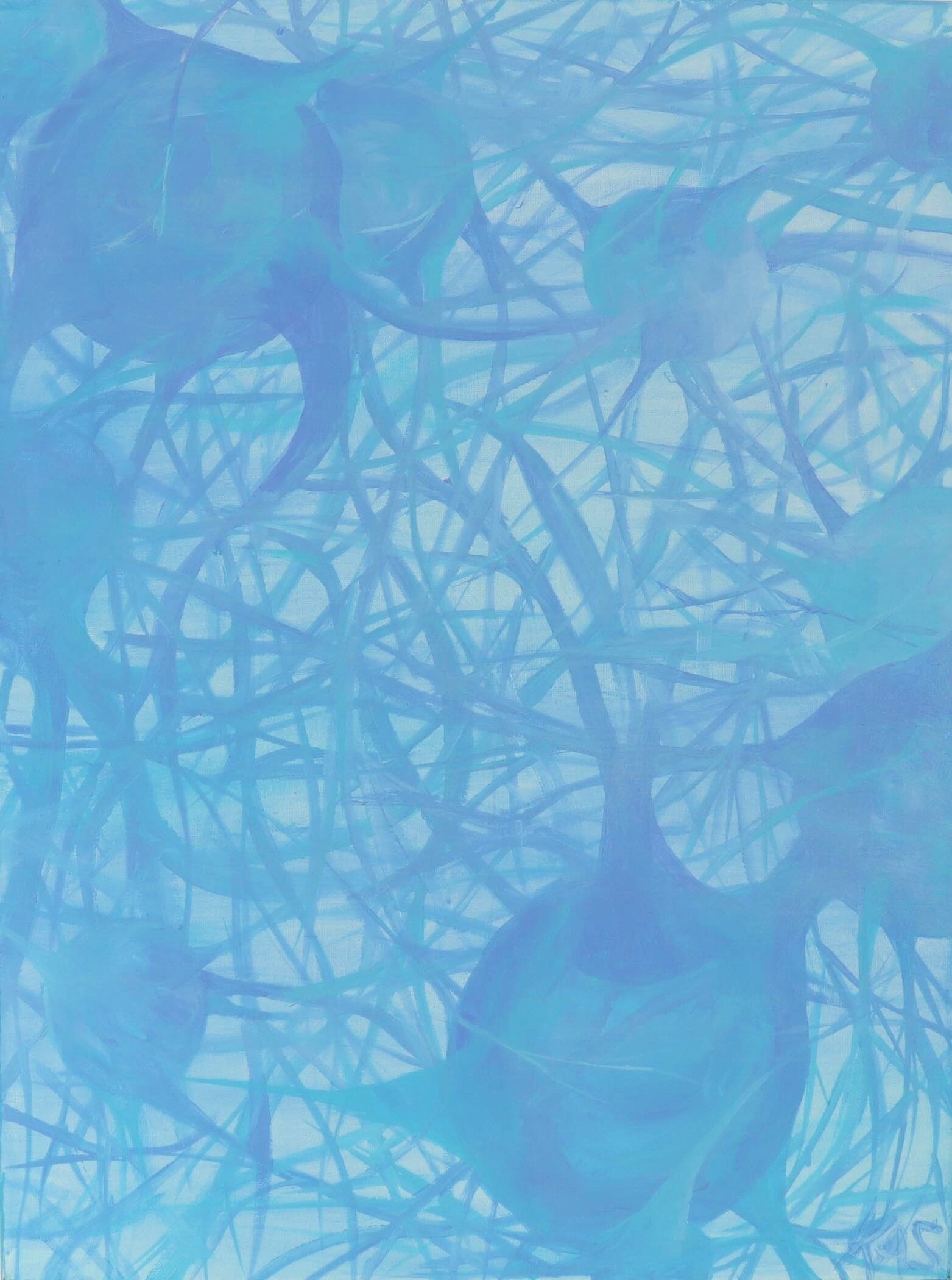
The clouded nature of the image represents the clinical symptom referred to as brain fog. Brain fog is described by patients with a number of conditions like chronic pain, after cancer treatment, and chronic fatigue syndrome. People with brain fog talk about difficulty concentrating and thinking.
24x30” Acrylic painting
This painting shows a variety of immune cells that would be found in inflamed joints with Rheumatoid Arthritis. Rheumatoid Arthritis is a disease that is characterized by joint inflammation and is associated with pain and stiffness. If untreated people get significant joint damage and deformities. Thanks to great science over the last 20 years, this is rarely observed. Known as an autoimmune disease, rheumatoid arthritis is the result of an immune system attacking its own tissues.
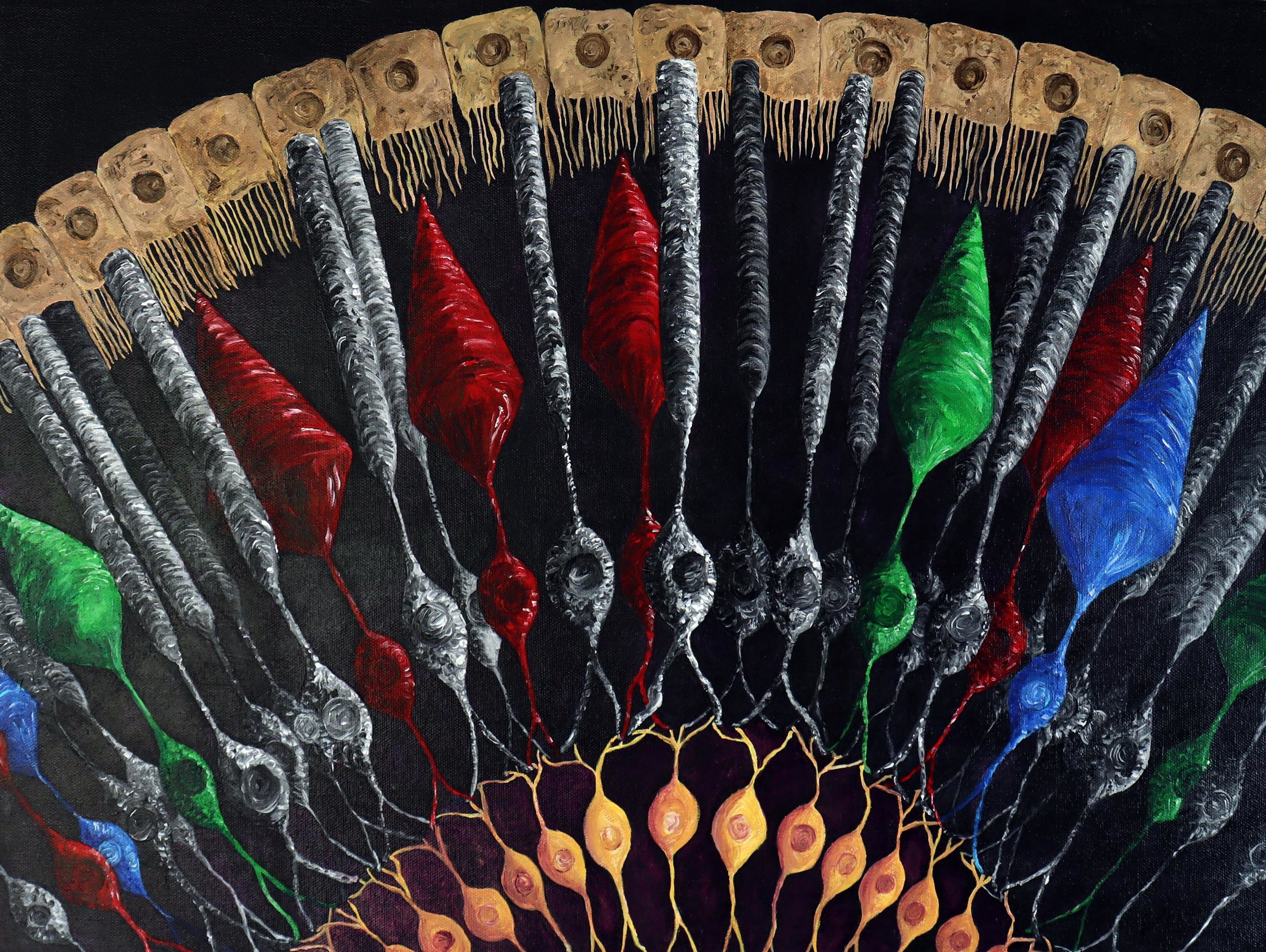
16x20” Acrylic Painting
Rods and Cones are specialized neurons found in the retina that are responsible for vision. These photoreceptor cells convert light to electrical signals to send information to the occipital lobe in the brain so that we can see. Rods, in black and white, are the most numerous and are activated by low levels of light. Cones, depicted in colors, are responsible for color vision. The signals from the rods and cones are transmitted through bipolar cells, in yellow at the bottom of the picture, to the optic nerve. Pigmented epithelial cells, top of the image, provide structure and nourishment to the photoreceptors.
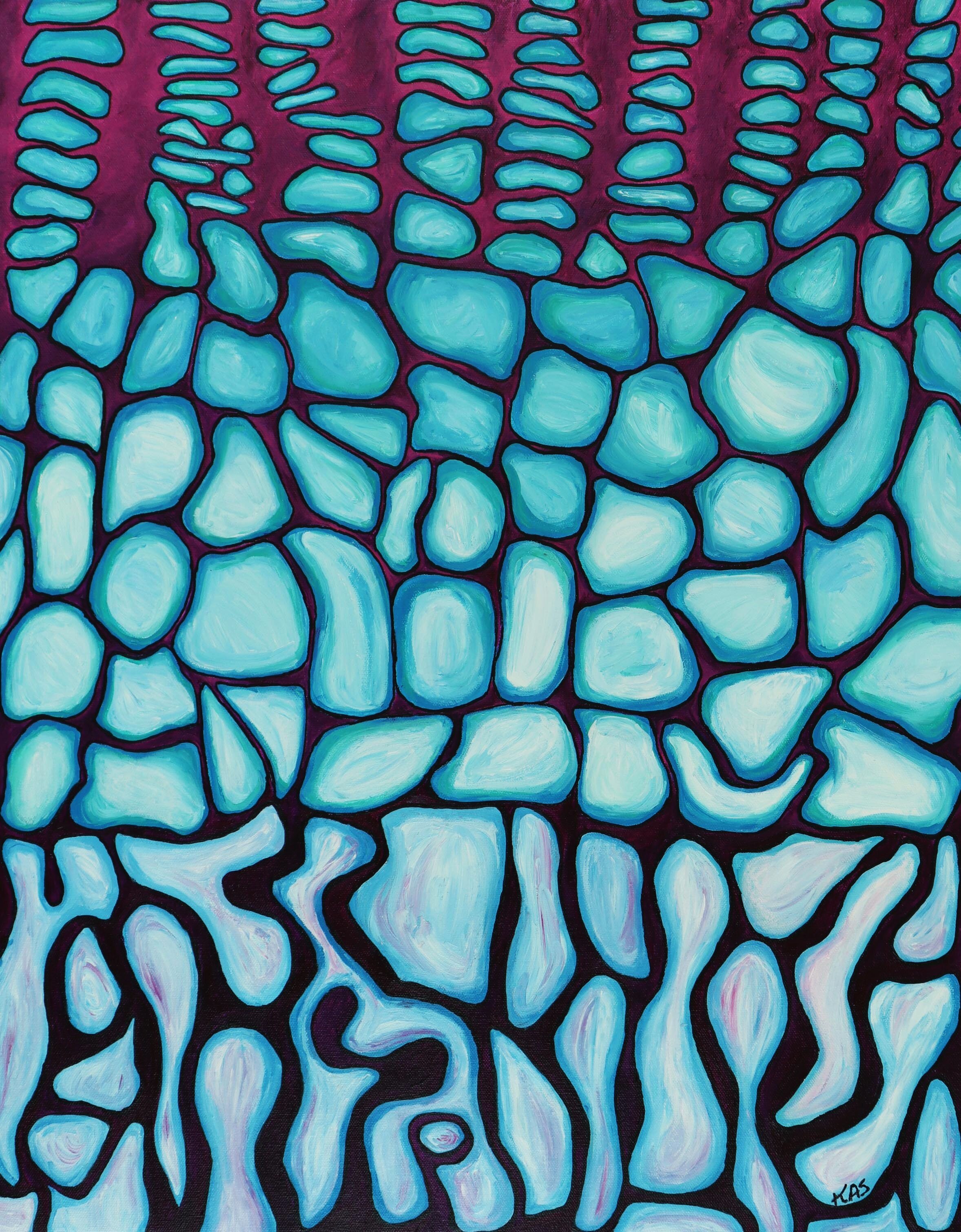
20x24” Acrylic painting
The growth plate, also known as the epiphyseal plate, is the area of growing bone in children, particularly in the long bones of the body-legs and arms. Each bone has two growth plates at each end. The top of the painting shows cartilage cells (blue) that continue to grow and produce new cells.

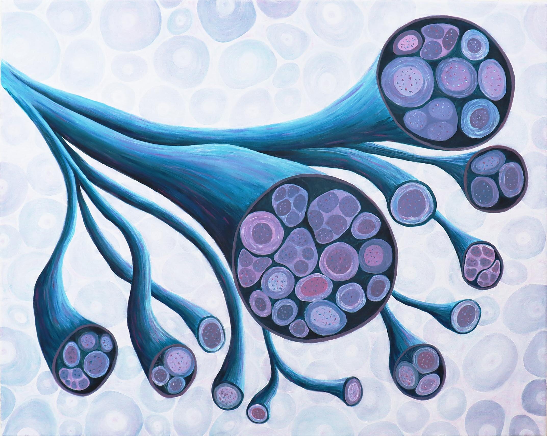
24x36” Acrylic Painting
This painting is a cross section of a nerve showing different sizes of nerve fibers. The largest ones are insulated by myelin, shown here as black rings. The largest nerve fibers, also called axons, send signals to our muscles to make them contract and move, and send signals from touch receptors so we can distinguish what we feel. The smallest, in red, orange and yellow, are thinly myelinated or unmyelinated and send pain signals to the spinal cord and brain. The unmyelinated axons congregate together in a Remak bundle.

24x36” Acrylic Painting
This painting shows loose connective tissue which surrounds all the tissue in the body. It is composed of collagen – yellow – and elastin – blue -which are produced by fibroblast cells – green.
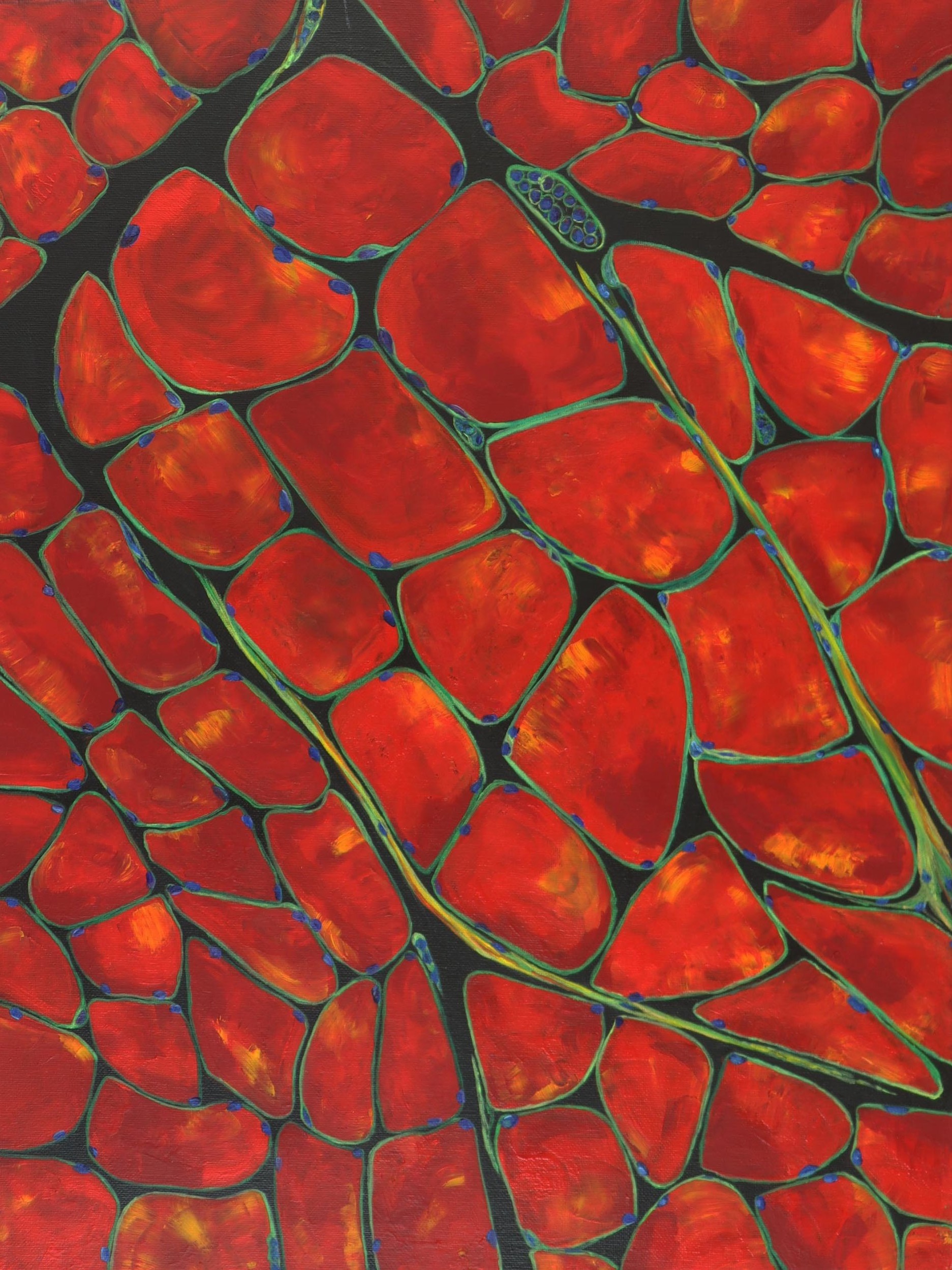
18x24” acrylic painting, SOLD
A muscle is composed of multiple bundles of cells called muscle fibers; these fibers generate the force to move our joints. Unlike other cells in the body, the muscle fibers have more than one nucleus, blue dots. Nerve fibers, in green, course around the muscle fibers, and include those that sense painful signals.
24×36″ Acrylic Painting
SOLD
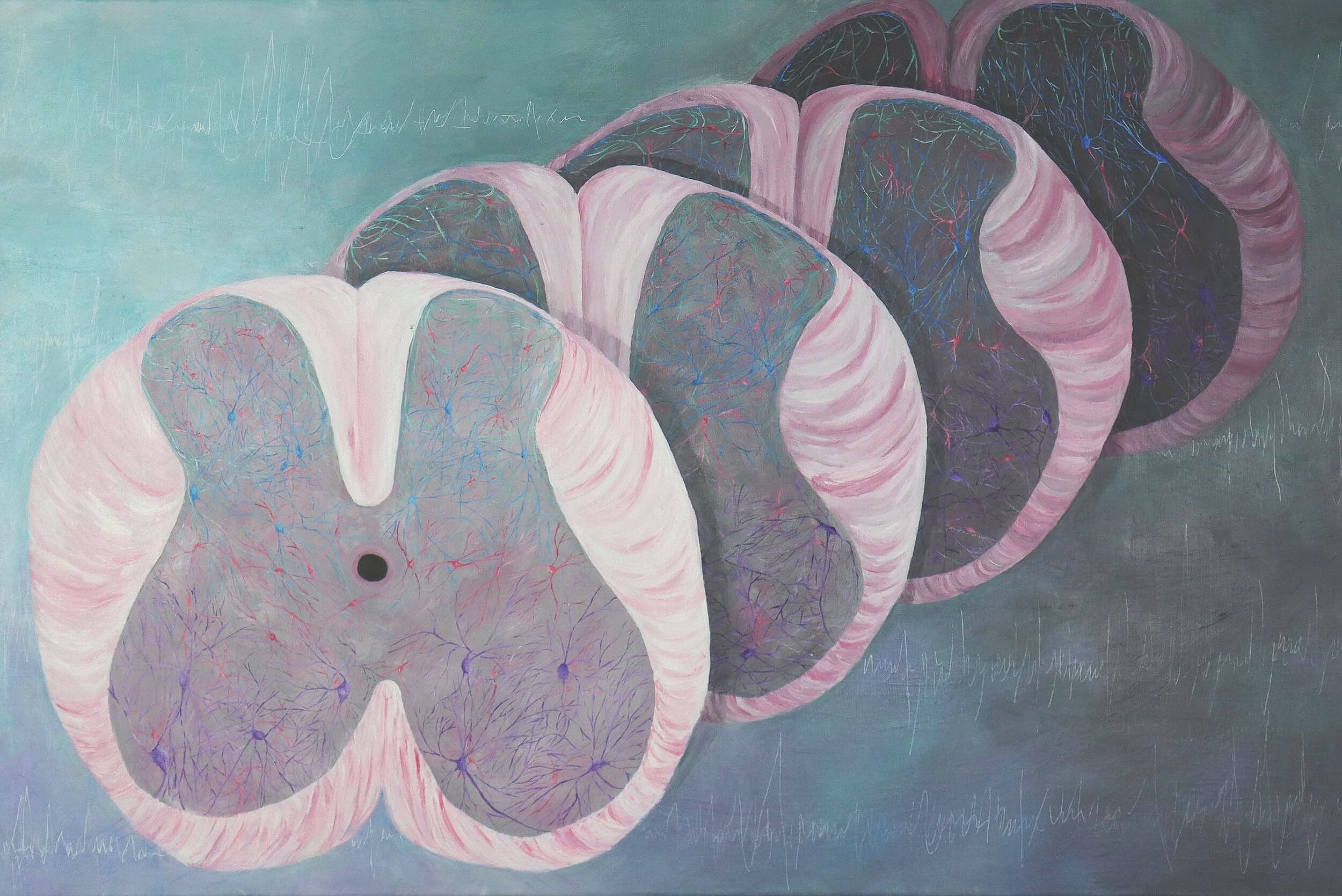
The spinal cord is divided into the dorsal horn-upper part, and the ventral horn-lower half. The dorsal horn receives input from sensory fibers in green. It sends input through interneurons, in pink, to spinothalamic tract cells, in blue. These cells are important for transmitting pain and temperature signals to the brain for perception of pain. The background shows action potentials which transmit electrical signals between neurons.
Cross sections of the midbrain showing the area involved In Parkinson’s disease - the substantial nigra. The dark neurons lose their pigment during Parkinson’s which results in loss of motor function.
8x10” Acrylic Painting
Sold
Neurons have intricate connections with other neurons through their dendrites and axons. It is through these connections that they send signals and messages to control nearly all aspects of the body including sensation, movement, breathing, learning, thoughts and emotions.
SOLD
18x24” acrylic painting
Alzheimer’s disease is associated with loss of memory and has a significant impact on the person. Alzheimer’s is associated with an abnormal build-up of proteins in and around brain cells. Beta-amyloid plaques form around neurons (yellow) and neurofibrillary tangles occur within neurons. Neurons stop functioning, lose connection with other neurons, and eventually die (dark purple).
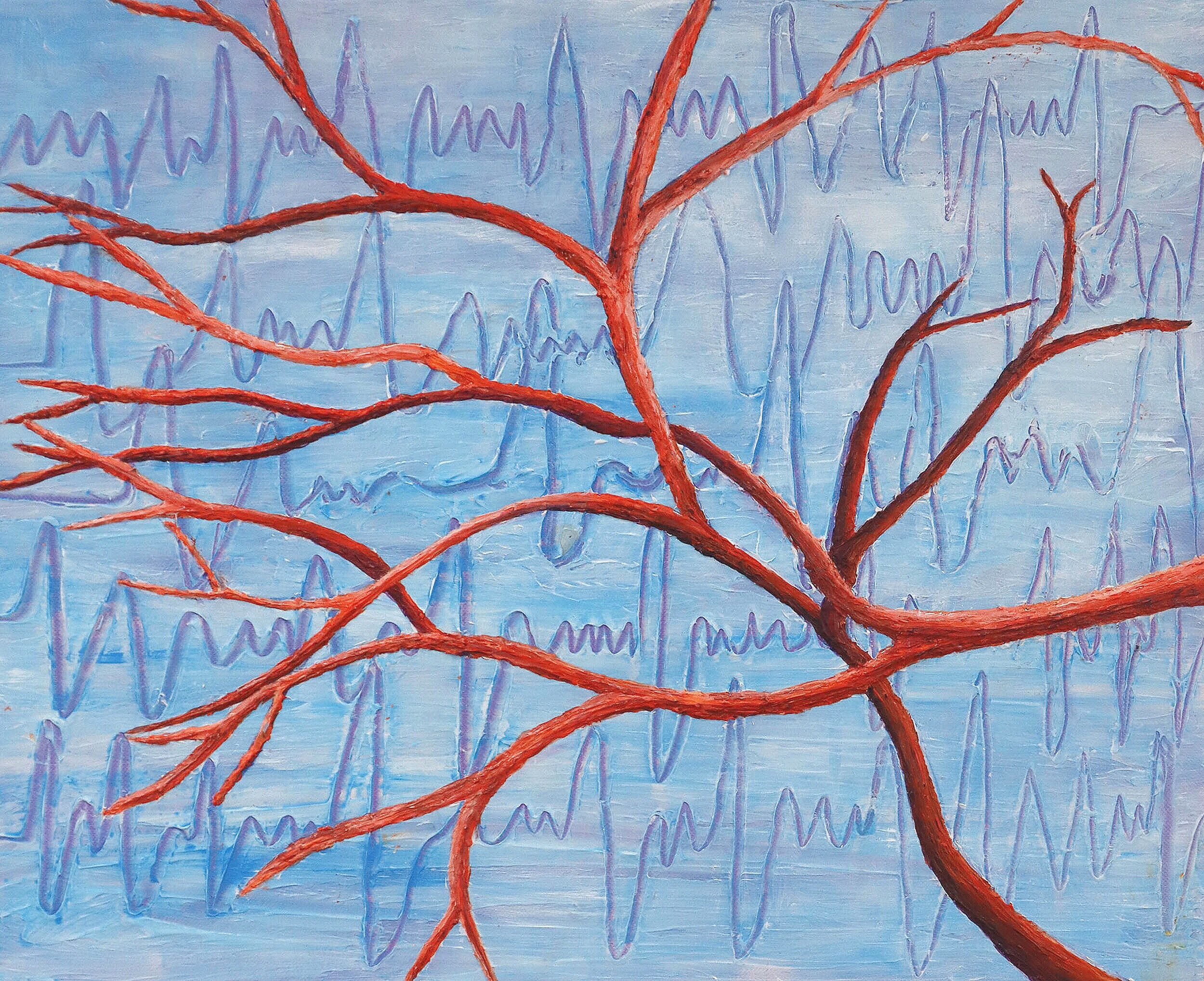
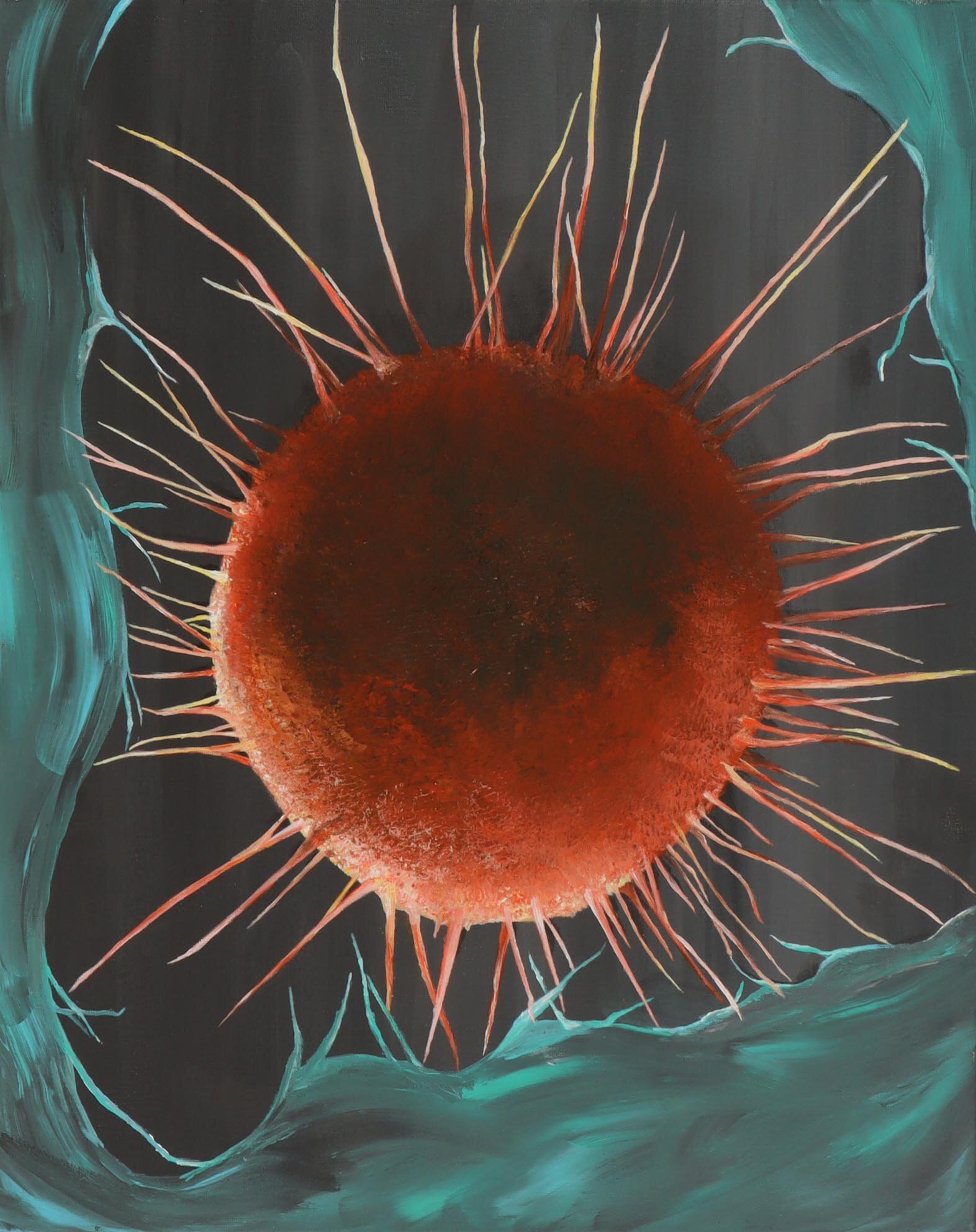
16x20” Original Acrylic Painting
Neurons transmit signals electrically through action potentials. When in pain, this sends up a red flag to the nervous system to warn against danger.
16x20” acrylic painting
Neurons transmit signals electrically through action potentials. When in pain, this sends up a red flag to the nervous system to warn against danger.
Trabecular bone, also referred to as spongy bone, is found at the end the long bones and contains bone marrow. Bone marrow produces red and white blood vessels. Blood vessels and nerves course throughout the marrow.